

Pulmonary Embolism: Symptoms, Causes, Diagnosis and Treatment
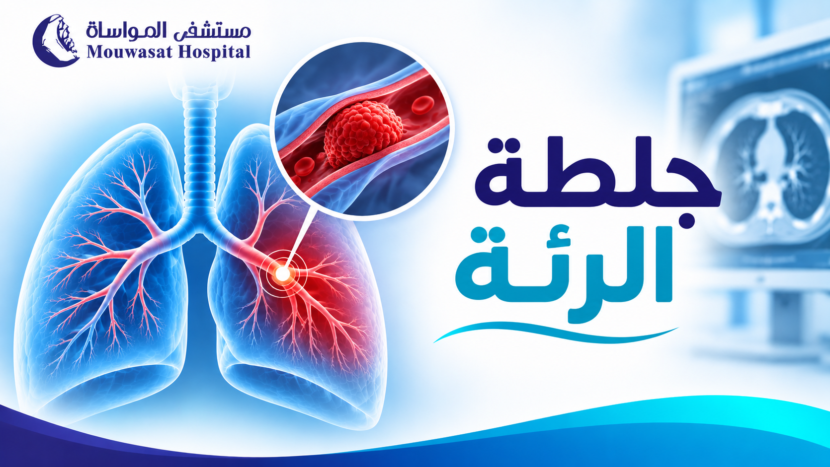
A pulmonary embolism (PE) occurs when a blood clot blocks one or more arteries in the lungs. In most cases, the clot does not originate in the lung itself. It forms in a deep vein—usually in the leg or pelvis—and travels through the bloodstream to the pulmonary arteries.[1]
The severity of PE ranges widely. Some patients are clinically stable and at relatively low short-term risk, while others develop severe hypoxemia, right-heart strain, hypotension, shock, or cardiac arrest.
- Sudden shortness of breath and pleuritic chest pain are common symptoms.
- Many pulmonary emboli originate from deep vein thrombosis in the legs or pelvis.
- An elevated D-dimer does not diagnose pulmonary embolism by itself.
- CT pulmonary angiography is a major imaging test when definitive imaging is required.
- PE severity depends on hemodynamic, cardiac and respiratory effects—not clot size alone.
- Anticoagulation is the foundation of treatment for most patients.
- Thrombolysis, catheter therapy or thrombectomy are reserved for selected higher-risk patients.
- Many people recover well, but follow-up is important for recurrent VTE and persistent symptoms.
- What is a pulmonary embolism?
- Where does the clot come from?
- Pulmonary embolism symptoms
- Early symptoms
- Can pulmonary embolism be mild?
- Emergency warning signs
- Causes and risk factors
- Travel and pulmonary embolism
- Cancer and pulmonary embolism
- How is PE diagnosed?
- What does D-dimer mean?
- CTPA and V/Q scanning
- How is severity determined?
- How is pulmonary embolism treated?
- Anticoagulation
- Thrombolysis
- Catheter treatment and thrombectomy
- Can pulmonary embolism be cured?
- How long does recovery take?
- Pulmonary embolism prognosis
- Can PE recur?
- CTEPD and CTEPH after PE
- Life after pulmonary embolism
- Pulmonary embolism care at Mouwasat Hospital
- Frequently asked questions
What Is a Pulmonary Embolism?
Pulmonary embolism is an acute obstruction of pulmonary arterial blood flow, most commonly caused by a blood clot that has travelled to the lungs.[1]
The obstruction can lower blood oxygen levels and increase pressure on the right ventricle, which must pump blood through the affected pulmonary circulation.
Severe PE therefore affects not only the lungs, but also the heart and systemic circulation.
Where Does a Pulmonary Embolism Come From?
Many pulmonary emboli begin as deep vein thrombosis (DVT) in the legs or pelvis.
A portion of the clot can detach and travel through the venous circulation and right side of the heart until it becomes lodged in a pulmonary artery.[1]
This is why clinicians ask about leg swelling, leg pain, previous DVT, surgery, immobility and other venous-thromboembolism risk factors.
What Are the Symptoms of Pulmonary Embolism?
Symptoms vary according to clot burden, cardiopulmonary reserve, and disease severity.
They can include:
- Sudden unexplained shortness of breath.
- Chest pain that may worsen with deep breathing.
- Faster-than-usual heart rate.
- Light-headedness.
- Fainting in more severe cases.
- Cough, sometimes with blood.
- Low oxygen levels.
Some pulmonary emboli are also discovered incidentally without obvious symptoms.[2]
What Are the Early Symptoms of Pulmonary Embolism?
A sudden change in breathing is one of the most recognizable presentations.
Patients may also develop new pleuritic chest pain, unexplained tachycardia, dizziness, or fainting.
However, PE can mimic pneumonia, myocardial infarction, asthma, musculoskeletal chest pain, and other conditions, so symptoms alone cannot establish the diagnosis.
Can Pulmonary Embolism Be Mild?
Yes, there are lower-risk and higher-risk forms of PE.
However, “mild PE” is not a safe self-diagnosis.
The 2026 AHA/ACC guideline introduced clinical categories A through E to describe severity more precisely, incorporating symptoms, clinical severity, right ventricular effects, biomarkers, respiratory status, and hemodynamic stability.[3]
When Is Pulmonary Embolism an Emergency?
- Sudden or severe shortness of breath.
- New chest pain with breathing difficulty.
- Fainting or altered consciousness.
- Severe oxygen desaturation or blue lips.
- Shock symptoms or persistent low blood pressure.
- Significant coughing of blood.
What Causes Pulmonary Embolism?
Risk increases when blood flow slows, clotting tendency increases, or blood-vessel injury occurs.
Important risk factors include:
- Previous DVT or PE.
- Major surgery.
- Prolonged immobility or hospitalization.
- Major trauma or fractures.
- Active cancer.
- Pregnancy and the postpartum period.
- Some estrogen-containing hormonal treatments.
- Obesity.
- Increasing age.
- Selected inherited or acquired thrombophilias.
Some episodes occur without a clear temporary trigger and are described as unprovoked PE.
Can Long-Distance Travel Cause Pulmonary Embolism?
Travel itself does not guarantee that a clot will develop, but prolonged sitting and immobility can contribute to venous stasis.
The absolute risk varies considerably according to individual factors such as previous VTE, recent surgery, active cancer, pregnancy, and other risks.
Does Cancer Increase the Risk of Pulmonary Embolism?
Yes. Malignancy can create a more pro-thrombotic state, while surgery, some treatments, hospitalization and immobility can further increase risk.
This is also why sudden dyspnea in a person with cancer should not automatically be assumed to come from the tumor itself.
How Is Pulmonary Embolism Diagnosed?
The diagnostic pathway begins with clinical assessment rather than automatically sending every patient for CT.
Depending on pretest probability, evaluation may include:
- D-dimer testing.
- CT pulmonary angiography.
- V/Q scanning in selected situations.
- ECG.
- Troponin or natriuretic peptides for risk assessment.
- Echocardiography for right-ventricular evaluation.
- Leg-vein ultrasound when DVT is suspected.

Does an Elevated D-dimer Mean I Have a Pulmonary Embolism?
No.
D-dimer can rise when blood clots are forming and breaking down, but it can also be elevated in infection, inflammation, pregnancy, cancer, after surgery, and with increasing age.
Its major value is helping clinicians exclude PE in appropriately selected patients with low or intermediate clinical probability.
The 2026 guideline supports age-adjusted D-dimer thresholds in appropriate older adults to safely reduce unnecessary imaging.[4]
When Are CTPA and V/Q Scans Used?
CT pulmonary angiography (CTPA) is the preferred confirmatory imaging test in many patients when imaging is indicated.[3]
A ventilation/perfusion scan can be useful when CTPA is unsuitable or when other clinical considerations favor an alternative.
This is why the diagnostic pathway uses clinical probability and D-dimer before imaging in selected patients rather than relying on CT for everyone.
How Do Doctors Determine How Dangerous a PE Is?
Clot size alone is not enough.
The 2026 AHA/ACC categories range from A to E:
| Category | Simplified description |
|---|---|
| A | Incidental or asymptomatic PE. |
| B | Symptomatic but lower clinical severity. |
| C | Higher clinical severity and/or evidence of cardiac strain or biomarker elevation. |
| D | Incipient cardiopulmonary failure. |
| E | Cardiopulmonary failure, including persistent hypotension, shock, or cardiac arrest. |
The classification guides disposition, monitoring, and treatment intensity.[3]
How Is Pulmonary Embolism Treated?
Treatment is risk-based.
Options may include:
- Anticoagulation.
- Oxygen and hemodynamic support when required.
- Systemic thrombolysis for selected higher-risk cases.
- Catheter-directed thrombolysis.
- Mechanical thrombectomy.
- Surgical embolectomy in selected situations.
Some appropriately selected lower-risk patients can be managed as outpatients or discharged early, whereas patients with elevated clinical severity, right-heart dysfunction or cardiopulmonary failure require hospital treatment.[3]
What Is the Role of Anticoagulation?
Anticoagulation is the foundation of treatment for most PE.
These medicines reduce the formation of new clots and prevent the existing clot from enlarging while the body's own processes gradually resolve the thrombus.
Treatment duration depends on whether PE was caused by a temporary risk factor, whether persistent risks remain, recurrence risk, and bleeding risk.
Patients should never start, stop, or change anticoagulation without medical supervision.
When Is Thrombolysis Used?
Thrombolytic medicines actively accelerate clot breakdown but can also substantially increase bleeding risk.
They are therefore not routine therapy for every PE and are used primarily when the expected benefit outweighs bleeding risk, particularly in patients with severe hemodynamic compromise.[3]
When Are Catheter Therapy or Mechanical Thrombectomy Used?
Catheter-based treatment can deliver clot-dissolving therapy locally or mechanically remove thrombus from the pulmonary circulation in selected patients.
The 2026 guideline considers advanced therapies—including catheter thrombolysis, mechanical thrombectomy and surgical embolectomy—particularly relevant to patients with severe or evolving cardiopulmonary failure.[3]
The Interventional Radiology Department at Mouwasat Hospital lists thrombolysis for vascular clots including DVT and pulmonary embolism among its vascular interventions.
Mouwasat has also published clinical cases illustrating the use of catheter intervention in severe PE:
- At Mouwasat Hospital Khobar, a patient with severe acute PE underwent therapeutic catheterization to remove pulmonary arterial thrombus.
- At Mouwasat Hospital Madinah, a patient with massive PE and cardiac arrest received thrombolysis followed by catheter thrombectomy after circulation was restored.
These cases illustrate what can be done in selected high-risk patients; they do not mean catheter intervention is required for every PE.
Can You Fully Recover From a Pulmonary Embolism?
Many people recover well after prompt diagnosis and appropriate treatment.
Recovery involves more than surviving the acute event. It also includes:
- Cardiopulmonary stabilization.
- Preventing recurrent thrombosis.
- Regaining physical capacity.
- Monitoring persistent breathlessness.
- Identifying chronic thromboembolic disease if it develops.
How Long Does Recovery From Pulmonary Embolism Take?
There is no universal recovery period.
Recovery depends on severity, right-heart involvement, oxygen requirements, clot burden, DVT, comorbid illness and complications.
The 2026 guideline recommends asking about PE-related symptoms and functional limitation at every visit for at least one year, helping identify chronic thromboembolic pulmonary disease and other causes of persistent dyspnea.[5]
What Is the Life Expectancy After Pulmonary Embolism?
There is no single meaningful life-expectancy number for PE.
Outcomes differ according to:
- Hemodynamic stability.
- Right ventricular strain.
- Speed of diagnosis and treatment.
- Active cancer and other comorbidities.
- Recurrent VTE risk.
- Treatment complications.
Many patients recover and return to their usual lives, while PE associated with shock or cardiac arrest carries substantially greater short-term risk.
Can Pulmonary Embolism Come Back?
Yes. Recurrence risk depends heavily on the reason the first clot developed.
A PE following a temporary major trigger such as surgery is different from PE associated with active cancer, persistent thrombophilia, previous VTE, or no identifiable reversible trigger.
This distinction helps determine the duration of anticoagulation and follow-up strategy.
What Are CTEPD and CTEPH?
Most patients do not develop chronic pulmonary vascular disease after PE, but persistent thromboembolic obstruction can cause long-term symptoms in a minority.
Chronic Thromboembolic Pulmonary Disease (CTEPD) describes chronic thromboembolic obstruction associated with persistent symptoms or exercise limitation.
If chronic thromboembolic disease produces pulmonary hypertension, it is called Chronic Thromboembolic Pulmonary Hypertension (CTEPH).
Persistent shortness of breath after PE therefore deserves evaluation rather than being automatically attributed to slow recovery.
Life After Pulmonary Embolism
Once medically stable, patients are generally encouraged to return gradually to appropriate physical activity rather than remain unnecessarily immobile.
Travel, exercise and work should be individualized according to disease severity, anticoagulation, bleeding risk and overall health.
Important follow-up points include:
- Taking anticoagulation exactly as prescribed.
- Not stopping treatment independently.
- Watching for bleeding.
- Reporting recurrent breathlessness or leg swelling.
- Keeping follow-up appointments.
If breathlessness has developed gradually over months rather than suddenly, different lung diseases may need consideration. Read Pulmonary Fibrosis: Symptoms, Causes, Diagnosis and Treatment for one important chronic cause of progressive dyspnea.

Pulmonary Embolism Care at Mouwasat Hospital
Management of severe PE can involve emergency medicine, cardiology, intensive care, interventional radiology and other specialties depending on the patient's condition.
The Mouwasat Interventional Radiology Department provides minimally invasive vascular interventions and lists thrombolysis for DVT and pulmonary embolism among its services.[6]
Published Mouwasat cases also demonstrate multidisciplinary management of massive PE involving emergency stabilization, intensive care, thrombolytic therapy and catheter thrombectomy when clinically appropriate.[7][8]
Frequently Asked Questions
What is usually the first symptom of pulmonary embolism?
Sudden unexplained shortness of breath is common, often with pleuritic chest pain, tachycardia, dizziness, or fainting. No one symptom confirms PE.
Can pulmonary embolism be fatal?
Yes, particularly when it causes shock or cardiac arrest. However, many patients recover well when PE is diagnosed and treated promptly.
Can pulmonary embolism be completely cured?
Many patients make an excellent functional recovery, although some require longer follow-up because of persistent symptoms or recurrent clot risk.
Does PE always start suddenly?
Symptoms often begin abruptly but can evolve over hours or days, and some pulmonary emboli are found incidentally.
Does a high D-dimer prove PE?
No. D-dimer is nonspecific and can be elevated in many other conditions.
Can a chest X-ray diagnose PE?
No. A chest X-ray may help identify alternative diagnoses but cannot reliably confirm or exclude pulmonary embolism.
Does every pulmonary embolism require intensive care?
No. Treatment location depends on clinical risk. Selected lower-risk patients may even be managed outside the hospital, while unstable cases require intensive monitoring.
Does every PE need thrombolysis?
No. Most patients are treated primarily with anticoagulation. Thrombolysis and advanced procedures are reserved for selected higher-risk situations.
Is thrombectomy better than anticoagulation?
They serve different purposes and are not interchangeable choices for every patient. Catheter thrombectomy is used selectively, while anticoagulation remains standard treatment for most PE.
Is pulmonary embolism the same as a heart attack?
No. PE usually involves a venous clot blocking a pulmonary artery. A heart attack typically involves obstruction of a coronary artery supplying the heart muscle.
Final Thoughts: PE Severity Is Determined by Its Effect on the Patient, Not Clot Size Alone
Pulmonary embolism ranges from low-risk stable disease to severe cardiopulmonary failure.
Appropriate management therefore depends on:
- Clinical probability.
- Oxygen and blood pressure.
- Right ventricular effects.
- Laboratory and imaging findings.
- Underlying disease and recurrent-clot risk.
With early diagnosis and appropriate risk-based treatment, many patients recover well. Follow-up after the acute event remains important for recurrent VTE, persistent dyspnea and chronic thromboembolic disease.
Medical disclaimer: Suspected acute pulmonary embolism requires urgent medical assessment. This article is educational and should not be used to diagnose PE or determine anticoagulant choice or duration.
Medical References
- American Heart Association — First AHA/ACC Acute Pulmonary Embolism Guideline
- AHA — Key Patient Messages for Acute Pulmonary Embolism
- 2026 AHA/ACC Guideline for Acute Pulmonary Embolism
- AHA — Acute PE Diagnostic Recommendations
- AHA — Top Things to Know: Acute Pulmonary Embolism
- Mouwasat Hospital — Interventional Radiology
- Mouwasat Hospital — Acute Pulmonary Embolism Case
- Mouwasat Hospital — Massive Pulmonary Embolism Treated in Madinah