How can i help you?
Latest Blogs
Explore All



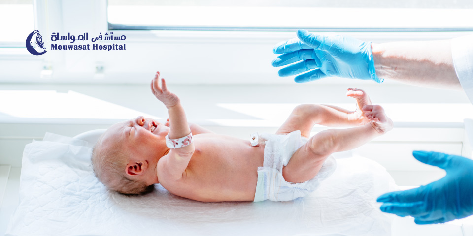

Children may experience urinary or genital problems that require specialized medical intervention. Some conditions are simple and can be treated by a pediatrician, but others need a pediatric urology surgeon to ensure accurate diagnosis and proper treatment.
In this article, we review the most common urinary and genital disorders in children, their causes, symptoms, and treatment options.
Pediatric urology surgery is a highly specialized branch of pediatric surgery focused on diagnosing, treating, and rehabilitating congenital and acquired disorders of the urinary and genital systems in children and adolescents.
Statistics indicate that urinary tract diseases in children account for 10% to 20% of all urological conditions, and approximately 75% of pediatric surgeries are urological, highlighting the importance of having an expert team and advanced techniques for optimal outcomes.
Children may suffer from urinary or genital issues, some requiring immediate medical attention or even surgery. Parents should pay attention to symptoms, especially in infants who cannot express pain.
The first step is usually visiting a pediatrician, but in complex cases or those unresponsive to treatment, the doctor may refer you to a pediatric urology specialist for a more accurate evaluation and tailored treatment plan.
Children may face various urinary tract and genital issues affecting normal urination or genital development. These problems fall into two main categories: urinary system disorders and genital system disorders.
A condition where urine flows backward from the bladder into the ureters and kidneys instead of exiting normally, due to a malfunctioning one-way valve.
Common Symptoms of VUR:
This condition increases the risk of recurrent urinary tract infections and may lead to kidney damage or failure if untreated.
Surgical repair between 6–12 months to reposition the urethral opening and correct curvature.
Al Mouwasat Hospital is among the leading centers for pediatric urology surgery, offering comprehensive care for complex and critical cases according to international standards. Our services include:
Our Pediatric Surgery Department provides integrated surgical care for infants, children, and adolescents, focusing on the highest safety and quality standards. Key features include:
Choosing Al Mouwasat Hospital means ensuring your child receives specialized, safe, and efficient care, making us the ideal destination for pediatric urology surgeries in the Kingdom
A mammogram is one of the main tools used for breast cancer screening because it can detect breast changes before they become large enough to feel or cause noticeable symptoms. However, mammography is not used for exactly the same purpose in every patient.
There is an important difference between a screening mammogram, which is performed in people without breast symptoms, and a diagnostic mammogram, which is used when there is a lump, nipple discharge, skin change or another finding that needs closer evaluation. [1] [2]
In Saudi Arabia, the Ministry of Health states that mammography is used to screen women aged 40 to 69 years. Women at higher risk may need an earlier or more intensive screening plan based on individual medical assessment. [1]
A mammogram is a low-dose X-ray image of the breast. It can reveal small changes that may not yet be detectable by touch. [2]
Mammography has two main uses:
The Saudi Ministry of Health states that mammography can help identify breast cancer at an early stage before a lesion becomes large enough to be felt. [2]
Saudi Ministry of Health early-detection guidance uses mammography for breast cancer screening in women aged 40 to 69 years. [1]
For a woman around age 40 who has no symptoms and is not known to be at high risk, this is a good time to discuss an appropriate screening schedule with her healthcare provider.
There is no single worldwide interval that applies to every woman. Frequency can depend on age, breast cancer risk, medical history, previous imaging findings and local recommendations.
The safest approach is to follow a screening schedule based on your individual situation rather than selecting an interval from a generic online rule.
| Screening mammogram | Diagnostic mammogram |
|---|---|
| Used when there are no breast symptoms. | Used when there is a symptom or abnormal finding to investigate. |
| Aims to find cancer before symptoms develop. | Aims to study a specific area or concern in greater detail. |
| Usually includes standard views of each breast. | May involve additional views, magnification or other imaging. |
| Part of routine early detection. | Part of a diagnostic work-up and may be combined with ultrasound or other tests. |
Saudi Ministry of Health guidance describes diagnostic mammography as the examination used when a woman has breast symptoms or when a previous mammogram shows a finding requiring further evaluation. [2]
If you have already noticed a change, see: early signs of breast cancer: changes you shouldn't ignore.

Being younger than 40 does not mean mammography is never appropriate. It means that routine population screening is not used in exactly the same way as it is in the main screening age group.
A younger woman with a new breast lump, another breast symptom, a strong family history or other risk factors may need imaging. Depending on the situation, the clinician may use ultrasound, mammography, MRI or a combination of tests.
The more useful question is therefore not simply, “Am I old enough for a mammogram?” but: “Am I symptom-free and considering screening, or do I have a breast problem that needs diagnosis?”
Some women need a personalised screening strategy because their breast cancer risk is significantly higher than average.
Important factors can include:
Saudi guidance notes that breast MRI may be used for women carrying certain genetic mutations associated with increased breast cancer risk. [1]
The American Cancer Society recommends annual mammography plus breast MRI for some women at high risk, typically beginning around age 30, depending on the individual risk profile. [4]
During the examination, you stand in front of the mammography machine. One breast is placed on a special plate and compressed briefly by another plate while an X-ray image is taken.
In a standard screening mammogram, two different views are usually taken of each breast, and the process is then repeated on the other side. [2]
Compression helps:
Saudi Ministry of Health guidance notes that the pressure lasts for a few seconds and does not harm the breast. [2]
Some women feel pressure or discomfort, and some may experience brief pain, particularly when the breasts are already tender.
The compression itself lasts only a short time for each image. The American Cancer Society suggests scheduling the test when the breasts are less swollen or tender if this is an issue for you. [6]
Mammography uses ionising X-rays, so it does involve radiation exposure. However, the radiation dose is low.
The FDA states that the risk of harm from mammography radiation is very small compared with the benefits of early breast cancer detection. [5]
Avoiding an appropriate mammogram because of the belief that the examination itself is more dangerous than an undetected breast cancer therefore does not reflect the balance of current evidence.
There is no evidence that the brief compression used during a mammogram spreads breast cancer. Compression is part of the imaging technique and helps produce a clearer image.
These preparation steps are recommended by the FDA and American Cancer Society to improve image quality and make the examination more comfortable. [5] [6]
Neither test is universally “better” because they are used for different purposes.
Mammography is the main imaging method used for population breast cancer screening, while ultrasound can help assess a specific lump or area and determine whether a mass is solid or fluid-filled. Depending on age, symptoms and breast characteristics, the two tests may be used together. [7]
Read the dedicated guide: mammogram vs breast ultrasound: when is each test used?.

An abnormal mammogram or a request for additional images does not automatically mean breast cancer.
A radiologist may request additional mammographic views, magnification, ultrasound or another examination because an area needs to be assessed more closely.
Breast imaging reports commonly use a standard system called BI-RADS to describe findings and recommend the next step.
See the upcoming guide: mammogram results and BI-RADS: what do categories 0 to 6 mean?.
No screening test is 100% accurate.
Some breast cancers may not be clearly visible on mammography, and mammographic sensitivity can be affected by several factors, particularly breast density. Saudi Ministry of Health information also notes that breast density can influence how well a mammogram detects cancer. [8]
A previous normal mammogram therefore should not be used as a reason to ignore a new breast lump or other new symptom.
No.
Being familiar with the usual appearance and feel of your breasts can help you notice a new change, but it cannot detect every small cancer that may be visible on a mammogram before it becomes palpable.
Not feeling a lump should therefore not be used as a reason to skip recommended breast cancer screening.
| Situation | Typical next step |
|---|---|
| Age 40 or older, no symptoms, average risk | Discuss an appropriate mammography screening schedule based on Saudi guidance and individual assessment. |
| New lump, nipple discharge, skin or nipple change | Diagnostic medical assessment rather than waiting for routine screening. |
| Under 40 with a new breast symptom | Your clinician selects the most appropriate imaging based on age and symptoms. |
| High inherited or family risk | An individualised plan may begin earlier and may include MRI with mammography. |
| Abnormal mammogram result | Additional imaging or other tests as recommended rather than assuming that cancer is present. |
Mammography for breast cancer screening is an important tool for early detection. Saudi Ministry of Health guidance uses mammography to screen women aged 40 to 69, but women at high risk may require a different strategy.
The most important distinction is between screening and diagnosis. If you have no symptoms, mammography may detect breast cancer before you can feel it. If you already have a lump, discharge, nipple change or another new symptom, the appropriate pathway is a diagnostic evaluation rather than simply waiting for routine screening.
Mammography also uses a low radiation dose, and the small radiation risk is outweighed by the benefits of appropriate early detection.
For a broader overview, see: breast cancer: from early signs to diagnosis, treatment and screening.
For specialist evaluation and treatment, visit Oncology at Mouwasat Hospital.
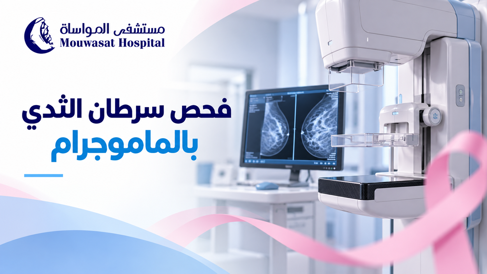

A new breast lump is one of the best-known early signs of breast cancer, but it is not the only possible sign. Some breast cancers can cause skin, nipple or breast-shape changes, while others may cause no noticeable symptoms at an early stage.
It is equally important to remember that a breast lump or change does not automatically mean cancer. Many breast changes are caused by benign conditions, hormonal changes, cysts or infections. A new or unusual change should, however, be medically assessed so its cause can be identified. [1] [3]
Symptoms vary from person to person, and some people have no symptoms at all in the early stages.
According to the Saudi Ministry of Health and CDC, breast changes that deserve attention include:
These symptoms can also occur in conditions that are not cancer. A new or unusual change should nevertheless be checked by a healthcare professional. [1] [2]
No. A new breast lump is the most common symptom, but breast cancer may first appear as a skin or nipple change, breast swelling, a change in shape, or an abnormal finding on imaging.
Mammography can also detect some breast cancers before they become large enough to be felt, so the absence of a lump does not remove the need for appropriate screening. [3]
To learn when screening is recommended, see: breast cancer screening and mammography: when should it start?.
No. A lump cannot be reliably diagnosed by touch alone.
A common belief is that a cancerous lump must always be hard, painless and fixed in place. Although many breast cancers may feel hard or irregular, the American Cancer Society notes that breast cancer lumps can also be round, soft, tender or painful. [3]
At the same time, most breast lumps are not cancer.
For more detail, see: breast lumps: when are they likely to be benign and when should they be checked?.
Sometimes the first noticeable change is in the skin rather than a lump.
Changes that should be assessed include:
The World Health Organization lists changes in breast size or shape, skin dimpling, redness and nipple or skin changes among possible symptoms of breast cancer. [4]
A new change in the nipple can sometimes be a symptom of breast cancer.
Changes may include:
Nipple discharge can also occur with benign conditions, so it is not a diagnosis of cancer by itself. Medical assessment can determine the cause. [2] [5]
Breast pain by itself is not usually a typical symptom of breast cancer. Most breast pain is caused by non-cancerous conditions such as hormonal changes, inflammation or problems involving nearby muscles and tissues.
However, new and persistent pain should not simply be ignored, particularly if it is accompanied by another breast change. The Saudi Ministry of Health includes new persistent breast or nipple pain among changes that warrant attention, while the NHS notes that breast pain alone is not usually a breast cancer symptom. [1] [5]
Yes. A new lump or thickening in the armpit can be a sign that needs medical assessment.
Lymph nodes in the armpit commonly enlarge because of infection or inflammation, but breast cancer can also spread to nearby lymph nodes. In some cases, enlarged lymph nodes may be noticed before a breast tumour is large enough to feel. [2] [3]
Breast redness and swelling can have many causes. Mastitis, for example, can cause redness, warmth, pain and a firm area in the breast, especially during breastfeeding. [6]
A less common form of breast cancer called inflammatory breast cancer can present differently from the typical breast lump. Possible changes include:
Rapid or unusual redness and swelling that does not improve as expected should therefore be assessed, particularly when there is no clear explanation.
Not every breast change is an emergency, but new, persistent or unusual changes should be medically assessed.
| Change | What to do |
|---|---|
| New breast or armpit lump | Arrange a medical assessment rather than trying to diagnose it by touch. |
| New change in breast size or shape | Have it checked if it is unusual for you or persists. |
| New nipple inversion or skin change | Seek medical assessment to identify the cause. |
| Bloody or unusual nipple discharge | Arrange a medical assessment. |
| New persistent breast pain | Have it assessed if it does not settle or occurs with another breast change. |
| Rapid redness, swelling or orange-peel skin | Do not delay assessment, particularly when there is no clear explanation. |
The CDC recommends medical assessment for breast signs or symptoms while emphasising that many such changes are caused by conditions other than cancer. [2]
No. This is important to remember.
The Saudi Ministry of Health and American Cancer Society note that breast changes can be caused by many non-cancerous conditions, including:
Because symptoms can overlap, the purpose of seeing a healthcare professional is not to assume the worst. It is to identify the cause rather than guess. [1] [3]
Breast cancer does not usually develop overnight, but a woman may notice a sign suddenly—for example, finding a lump while showering or noticing a skin change in the mirror.
Cancer can also be present before it becomes visible or large enough to feel. In many cases, “sudden” refers to when the change was first noticed rather than when the disease began.
| Symptoms | Screening |
|---|---|
| A breast change has already been noticed. | Performed in people without symptoms according to age and risk. |
| The change may require diagnostic assessment. | Designed to detect cancer before symptoms appear. |
| Evaluation should not be delayed until the next routine screening date. | Usually follows a recommended mammography schedule based on individual risk. |
If you have a new lump or symptom, the appropriate next step may be a clinical examination and diagnostic imaging rather than simply waiting for your next routine mammogram.
If you have no symptoms, screening remains important because some breast cancers can be detected before any noticeable change develops. [1] [3]
Knowing how your breasts normally look and feel can help you notice a new change, but checking your breasts does not prevent cancer and should not replace recommended mammography or medical evaluation.
Breast awareness helps identify what is unusual for you; determining the cause requires professional assessment. [5]
Breast cancer can also occur in men, although it is much less common. Possible signs include a breast lump, nipple changes or discharge.
Read more in: breast cancer in men: signs that can easily be missed.
A new lump may be one of the first signs of breast cancer, but the earliest noticeable change may also involve the skin, nipple, breast shape, unusual discharge or the lymph nodes under the arm.
At the same time, most breast changes and lumps are not cancer. Finding a change does not mean you have breast cancer; it means that identifying the cause is better than trying to diagnose it from pain, appearance or touch alone.
Screening also remains important even when you feel well because some breast cancers can be detected before symptoms appear.
For a broader overview, see: breast cancer: from early signs to diagnosis, treatment and screening.
For specialist evaluation and care, visit Oncology at Mouwasat Hospital.
A new breast lump is one of the best-known early signs of breast cancer, but it is not the only possible sign. Some breast cancers can cause skin, nipple or breast-shape changes, while others may cause no noticeable symptoms at an early stage.
It is equally important to remember that a breast lump or change does not automatically mean cancer. Many breast changes are caused by benign conditions, hormonal changes, cysts or infections. A new or unusual change should, however, be medically assessed so its cause can be identified. [1] [3]
Symptoms vary from person to person, and some people have no symptoms at all in the early stages.
According to the Saudi Ministry of Health and CDC, breast changes that deserve attention include:
These symptoms can also occur in conditions that are not cancer. A new or unusual change should nevertheless be checked by a healthcare professional. [1] [2]
No. A new breast lump is the most common symptom, but breast cancer may first appear as a skin or nipple change, breast swelling, a change in shape, or an abnormal finding on imaging.
Mammography can also detect some breast cancers before they become large enough to be felt, so the absence of a lump does not remove the need for appropriate screening. [3]
To learn when screening is recommended, see: breast cancer screening and mammography: when should it start?.
No. A lump cannot be reliably diagnosed by touch alone.
A common belief is that a cancerous lump must always be hard, painless and fixed in place. Although many breast cancers may feel hard or irregular, the American Cancer Society notes that breast cancer lumps can also be round, soft, tender or painful. [3]
At the same time, most breast lumps are not cancer.
For more detail, see: breast lumps: when are they likely to be benign and when should they be checked?.
Sometimes the first noticeable change is in the skin rather than a lump.
Changes that should be assessed include:
The World Health Organization lists changes in breast size or shape, skin dimpling, redness and nipple or skin changes among possible symptoms of breast cancer. [4]
A new change in the nipple can sometimes be a symptom of breast cancer.
Changes may include:
Nipple discharge can also occur with benign conditions, so it is not a diagnosis of cancer by itself. Medical assessment can determine the cause. [2] [5]
Breast pain by itself is not usually a typical symptom of breast cancer. Most breast pain is caused by non-cancerous conditions such as hormonal changes, inflammation or problems involving nearby muscles and tissues.
However, new and persistent pain should not simply be ignored, particularly if it is accompanied by another breast change. The Saudi Ministry of Health includes new persistent breast or nipple pain among changes that warrant attention, while the NHS notes that breast pain alone is not usually a breast cancer symptom. [1] [5]
Yes. A new lump or thickening in the armpit can be a sign that needs medical assessment.
Lymph nodes in the armpit commonly enlarge because of infection or inflammation, but breast cancer can also spread to nearby lymph nodes. In some cases, enlarged lymph nodes may be noticed before a breast tumour is large enough to feel. [2] [3]
Breast redness and swelling can have many causes. Mastitis, for example, can cause redness, warmth, pain and a firm area in the breast, especially during breastfeeding. [6]
A less common form of breast cancer called inflammatory breast cancer can present differently from the typical breast lump. Possible changes include:
Rapid or unusual redness and swelling that does not improve as expected should therefore be assessed, particularly when there is no clear explanation.
Not every breast change is an emergency, but new, persistent or unusual changes should be medically assessed.
| Change | What to do |
|---|---|
| New breast or armpit lump | Arrange a medical assessment rather than trying to diagnose it by touch. |
| New change in breast size or shape | Have it checked if it is unusual for you or persists. |
| New nipple inversion or skin change | Seek medical assessment to identify the cause. |
| Bloody or unusual nipple discharge | Arrange a medical assessment. |
| New persistent breast pain | Have it assessed if it does not settle or occurs with another breast change. |
| Rapid redness, swelling or orange-peel skin | Do not delay assessment, particularly when there is no clear explanation. |
The CDC recommends medical assessment for breast signs or symptoms while emphasising that many such changes are caused by conditions other than cancer. [2]
No. This is important to remember.
The Saudi Ministry of Health and American Cancer Society note that breast changes can be caused by many non-cancerous conditions, including:
Because symptoms can overlap, the purpose of seeing a healthcare professional is not to assume the worst. It is to identify the cause rather than guess. [1] [3]
Breast cancer does not usually develop overnight, but a woman may notice a sign suddenly—for example, finding a lump while showering or noticing a skin change in the mirror.
Cancer can also be present before it becomes visible or large enough to feel. In many cases, “sudden” refers to when the change was first noticed rather than when the disease began.
| Symptoms | Screening |
|---|---|
| A breast change has already been noticed. | Performed in people without symptoms according to age and risk. |
| The change may require diagnostic assessment. | Designed to detect cancer before symptoms appear. |
| Evaluation should not be delayed until the next routine screening date. | Usually follows a recommended mammography schedule based on individual risk. |
If you have a new lump or symptom, the appropriate next step may be a clinical examination and diagnostic imaging rather than simply waiting for your next routine mammogram.
If you have no symptoms, screening remains important because some breast cancers can be detected before any noticeable change develops. [1] [3]
Knowing how your breasts normally look and feel can help you notice a new change, but checking your breasts does not prevent cancer and should not replace recommended mammography or medical evaluation.
Breast awareness helps identify what is unusual for you; determining the cause requires professional assessment. [5]
Breast cancer can also occur in men, although it is much less common. Possible signs include a breast lump, nipple changes or discharge.
Read more in: breast cancer in men: signs that can easily be missed.
A new lump may be one of the first signs of breast cancer, but the earliest noticeable change may also involve the skin, nipple, breast shape, unusual discharge or the lymph nodes under the arm.
At the same time, most breast changes and lumps are not cancer. Finding a change does not mean you have breast cancer; it means that identifying the cause is better than trying to diagnose it from pain, appearance or touch alone.
Screening also remains important even when you feel well because some breast cancers can be detected before symptoms appear.
For a broader overview, see: breast cancer: from early signs to diagnosis, treatment and screening.
For specialist evaluation and care, visit Oncology at Mouwasat Hospital.
A mammogram is one of the main tools used for breast cancer screening because it can detect breast changes before they become large enough to feel or cause noticeable symptoms. However, mammography is not used for exactly the same purpose in every patient.
There is an important difference between a screening mammogram, which is performed in people without breast symptoms, and a diagnostic mammogram, which is used when there is a lump, nipple discharge, skin change or another finding that needs closer evaluation. [1] [2]
In Saudi Arabia, the Ministry of Health states that mammography is used to screen women aged 40 to 69 years. Women at higher risk may need an earlier or more intensive screening plan based on individual medical assessment. [1]
A mammogram is a low-dose X-ray image of the breast. It can reveal small changes that may not yet be detectable by touch. [2]
Mammography has two main uses:
The Saudi Ministry of Health states that mammography can help identify breast cancer at an early stage before a lesion becomes large enough to be felt. [2]
Saudi Ministry of Health early-detection guidance uses mammography for breast cancer screening in women aged 40 to 69 years. [1]
For a woman around age 40 who has no symptoms and is not known to be at high risk, this is a good time to discuss an appropriate screening schedule with her healthcare provider.
There is no single worldwide interval that applies to every woman. Frequency can depend on age, breast cancer risk, medical history, previous imaging findings and local recommendations.
The safest approach is to follow a screening schedule based on your individual situation rather than selecting an interval from a generic online rule.
| Screening mammogram | Diagnostic mammogram |
|---|---|
| Used when there are no breast symptoms. | Used when there is a symptom or abnormal finding to investigate. |
| Aims to find cancer before symptoms develop. | Aims to study a specific area or concern in greater detail. |
| Usually includes standard views of each breast. | May involve additional views, magnification or other imaging. |
| Part of routine early detection. | Part of a diagnostic work-up and may be combined with ultrasound or other tests. |
Saudi Ministry of Health guidance describes diagnostic mammography as the examination used when a woman has breast symptoms or when a previous mammogram shows a finding requiring further evaluation. [2]
If you have already noticed a change, see: early signs of breast cancer: changes you shouldn't ignore.
Being younger than 40 does not mean mammography is never appropriate. It means that routine population screening is not used in exactly the same way as it is in the main screening age group.
A younger woman with a new breast lump, another breast symptom, a strong family history or other risk factors may need imaging. Depending on the situation, the clinician may use ultrasound, mammography, MRI or a combination of tests.
The more useful question is therefore not simply, “Am I old enough for a mammogram?” but: “Am I symptom-free and considering screening, or do I have a breast problem that needs diagnosis?”
Some women need a personalised screening strategy because their breast cancer risk is significantly higher than average.
Important factors can include:
Saudi guidance notes that breast MRI may be used for women carrying certain genetic mutations associated with increased breast cancer risk. [1]
The American Cancer Society recommends annual mammography plus breast MRI for some women at high risk, typically beginning around age 30, depending on the individual risk profile. [4]
During the examination, you stand in front of the mammography machine. One breast is placed on a special plate and compressed briefly by another plate while an X-ray image is taken.
In a standard screening mammogram, two different views are usually taken of each breast, and the process is then repeated on the other side. [2]
Compression helps:
Saudi Ministry of Health guidance notes that the pressure lasts for a few seconds and does not harm the breast. [2]
Some women feel pressure or discomfort, and some may experience brief pain, particularly when the breasts are already tender.
The compression itself lasts only a short time for each image. The American Cancer Society suggests scheduling the test when the breasts are less swollen or tender if this is an issue for you. [6]
Mammography uses ionising X-rays, so it does involve radiation exposure. However, the radiation dose is low.
The FDA states that the risk of harm from mammography radiation is very small compared with the benefits of early breast cancer detection. [5]
Avoiding an appropriate mammogram because of the belief that the examination itself is more dangerous than an undetected breast cancer therefore does not reflect the balance of current evidence.
There is no evidence that the brief compression used during a mammogram spreads breast cancer. Compression is part of the imaging technique and helps produce a clearer image.
These preparation steps are recommended by the FDA and American Cancer Society to improve image quality and make the examination more comfortable. [5] [6]
Neither test is universally “better” because they are used for different purposes.
Mammography is the main imaging method used for population breast cancer screening, while ultrasound can help assess a specific lump or area and determine whether a mass is solid or fluid-filled. Depending on age, symptoms and breast characteristics, the two tests may be used together. [7]
Read the dedicated guide: mammogram vs breast ultrasound: when is each test used?.
An abnormal mammogram or a request for additional images does not automatically mean breast cancer.
A radiologist may request additional mammographic views, magnification, ultrasound or another examination because an area needs to be assessed more closely.
Breast imaging reports commonly use a standard system called BI-RADS to describe findings and recommend the next step.
See the upcoming guide: mammogram results and BI-RADS: what do categories 0 to 6 mean?.
No screening test is 100% accurate.
Some breast cancers may not be clearly visible on mammography, and mammographic sensitivity can be affected by several factors, particularly breast density. Saudi Ministry of Health information also notes that breast density can influence how well a mammogram detects cancer. [8]
A previous normal mammogram therefore should not be used as a reason to ignore a new breast lump or other new symptom.
No.
Being familiar with the usual appearance and feel of your breasts can help you notice a new change, but it cannot detect every small cancer that may be visible on a mammogram before it becomes palpable.
Not feeling a lump should therefore not be used as a reason to skip recommended breast cancer screening.
| Situation | Typical next step |
|---|---|
| Age 40 or older, no symptoms, average risk | Discuss an appropriate mammography screening schedule based on Saudi guidance and individual assessment. |
| New lump, nipple discharge, skin or nipple change | Diagnostic medical assessment rather than waiting for routine screening. |
| Under 40 with a new breast symptom | Your clinician selects the most appropriate imaging based on age and symptoms. |
| High inherited or family risk | An individualised plan may begin earlier and may include MRI with mammography. |
| Abnormal mammogram result | Additional imaging or other tests as recommended rather than assuming that cancer is present. |
Mammography for breast cancer screening is an important tool for early detection. Saudi Ministry of Health guidance uses mammography to screen women aged 40 to 69, but women at high risk may require a different strategy.
The most important distinction is between screening and diagnosis. If you have no symptoms, mammography may detect breast cancer before you can feel it. If you already have a lump, discharge, nipple change or another new symptom, the appropriate pathway is a diagnostic evaluation rather than simply waiting for routine screening.
Mammography also uses a low radiation dose, and the small radiation risk is outweighed by the benefits of appropriate early detection.
For a broader overview, see: breast cancer: from early signs to diagnosis, treatment and screening.
For specialist evaluation and treatment, visit Oncology at Mouwasat Hospital.
Breast cancer is not a single disease with one presentation or one treatment. It is a group of cancers that develop when abnormal cells in breast tissue begin growing out of control. Most breast cancers begin in the milk ducts or milk-producing lobules. Some remain confined to their site of origin, while invasive cancers can grow into surrounding breast tissue and may spread to lymph nodes or other organs. [1]
Breast cancer is also not diagnosed simply by feeling a lump. Some cancers are detected on mammography before symptoms appear, while others first cause a change in the breast, skin or nipple. When imaging identifies a suspicious area, a biopsy is needed to determine whether the cells are cancerous. Further testing then helps establish the cancer's biological features and guide treatment. [2]
Breast cancer develops when cells in the breast begin to grow abnormally and uncontrollably. Most breast cancers start in the ducts that carry milk or in the lobules that produce it. [1]
Abnormal cells can initially remain confined to where they started, known as in situ disease. If they grow through surrounding tissue, the cancer becomes invasive. Some invasive cancers can reach nearby lymph nodes and later spread to distant organs. [1]
Yes. Breast cancer is overwhelmingly more common in women, but men can develop it as well. The World Health Organization estimates that approximately 0.5% to 1% of breast cancers occur in men. [1]
No. Breast cancers differ in where they begin, their microscopic characteristics, their biological behaviour and their response to treatment.
At a basic level, breast cancer may be described as:
After a biopsy, however, doctors also assess biological characteristics such as estrogen and progesterone receptors and HER2 because these findings can directly affect treatment choices. [3]
There is no single cause that explains every breast cancer. Having a risk factor does not mean that cancer will occur, and having no obvious risk factors does not mean that a woman is protected.
The World Health Organization reports that approximately 80% of breast cancers occur in women without specific identifiable risk factors other than sex and age. [1]
Factors associated with increased risk include:
This is why the absence of a family history should not be used to dismiss a new breast symptom or appropriate screening. [1]
Early breast cancer may cause no noticeable symptoms and can sometimes be detected by imaging before a woman can feel anything.
Possible signs include:
These signs do not diagnose breast cancer. Many are caused by benign conditions, but a new or unusual breast change should be medically assessed. [2]
Diagnosis is a pathway rather than a single test.
A woman may enter this pathway because screening detected an abnormality before symptoms developed, or because she noticed a breast lump, nipple change, discharge, skin change or another symptom.
Assessment may include a clinical breast examination, mammography, breast ultrasound and, when indicated, other imaging. If the finding remains suspicious, a biopsy is performed. [2]
No. Imaging can show that an area is suspicious and help doctors characterise it, but imaging alone cannot prove that the cells are cancerous.
Mammography, ultrasound, MRI or a combination may be used depending on the patient's age, symptoms, breast characteristics and level of risk.
When imaging identifies an area that needs further evaluation, a biopsy removes a small sample of tissue so that it can be examined in a laboratory.
The Saudi Ministry of Health identifies biopsy as the definitive method for diagnosing breast cancer. The tissue can also be tested to determine tumour characteristics, grade and receptor status. [2]
Being referred for a biopsy therefore does not mean that cancer has already been diagnosed. It means that imaging has identified a question that requires tissue analysis for a reliable answer.
Once cancer is confirmed, knowing its stage is not enough. Doctors also need to understand what is driving the tumour biologically.
| Marker | What it means | Why it matters |
|---|---|---|
| ER | Estrogen receptor. | ER-positive cancers may respond to endocrine therapy. |
| PR | Progesterone receptor. | Together with ER, it helps define hormone sensitivity. |
| HER2 | A protein that is present at high levels in some breast cancers. | HER2-positive cancers may be treated with HER2-targeted therapies. |
Triple-negative breast cancer does not express ER or PR and does not have HER2 overexpression, so its treatment strategy differs from hormone receptor-positive and HER2-positive disease. [1] [3]
| Information | Question it answers |
|---|---|
| Type / subtype | What kind of cancer is it and what are its biological features? |
| Stage | How far has the cancer spread within the breast, lymph nodes or elsewhere? |
| Grade | How abnormal do the cancer cells look and what may this suggest about their growth behaviour? |
Two patients can therefore have breast cancer at the same stage and still receive different treatment because the biology of their tumours is different.
Breast cancer is generally staged from 0 to IV. Modern staging considers more than tumour size. It incorporates tumour and lymph-node involvement, distant spread, tumour grade and biological markers such as ER, PR and HER2. [4]
| Stage | General meaning |
|---|---|
| 0 | Non-invasive disease such as ductal carcinoma in situ (DCIS). |
| I | Early invasive cancer that remains relatively limited. |
| II | A larger tumour and/or involvement of some nearby lymph nodes, depending on the specific findings. |
| III | Locally advanced disease involving nearby tissues and/or more extensive regional lymph nodes, without distant metastasis. |
| IV | Breast cancer that has spread to a distant organ. |
This table is only a simplified overview. Formal staging is more detailed and cannot be determined from tumour size or symptoms alone. [4]
There is no universal breast cancer treatment plan. Treatment depends on factors including:
For this reason, two patients with the same numerical stage can receive different treatment combinations. [3] [5]
Surgery may remove the tumour and a margin of surrounding tissue while preserving the rest of the breast, or may involve mastectomy in other cases. The choice depends on the tumour, its extent, the ability to use radiation therapy and individual clinical considerations.
Nearby lymph nodes may also need to be assessed, including sentinel lymph-node biopsy when appropriate. [1]
Radiation therapy targets a defined area with high-energy radiation to reduce the risk of cancer returning locally. It is commonly used after breast-conserving surgery and may also be recommended after mastectomy in selected situations. [3]
Chemotherapy is systemic treatment that reaches cancer cells through the bloodstream. It can be given before surgery, after surgery or as part of treatment for advanced disease, depending on the cancer's characteristics.
Hormone receptor-positive breast cancers may respond to medicines that block hormone signalling or reduce hormonal stimulation of the cancer. [1]
Targeted therapies act on specific abnormalities within cancer cells. HER2-positive breast cancer is an important example because treatments are available that specifically target the HER2 pathway.
Immunotherapy can be used in selected breast cancers based on tumour type, stage and relevant test results. It is not routinely appropriate for every person with breast cancer.
There is an important difference between early diagnosis and screening.
Saudi Ministry of Health guidance uses mammography for breast cancer screening in women aged 40 to 69 years. Women with higher inherited or medical risk may require a different strategy. [6]
Not every breast cancer can be prevented. Many risk factors cannot be changed, and cancer can occur even without a known family history.
However, some modifiable risks may be reduced through:
Risk reduction is not a guarantee of prevention and does not replace appropriate screening or evaluation of new symptoms. [1]
Yes. Many breast cancers can be treated successfully, particularly when they are detected and treated early. There is, however, no single survival or cure percentage that applies to every patient. [1]
Outcome depends on factors such as:
Stage IV breast cancer has spread to a distant part of the body. Treatment generally focuses on controlling the cancer, slowing its progression, relieving symptoms, prolonging life and maintaining quality of life, with treatment selected according to the tumour's biology and the individual patient's situation. [5]
Together, these answers provide a much more useful picture than the words “breast cancer” or a stage number alone.
Breast cancer is a group of diseases that differ in where they begin, their biological features, their extent and their response to treatment. It may be found because of a breast change or detected through screening before any symptoms develop.
Imaging helps assess suspicious findings, but biopsy confirms the diagnosis. Stage, grade, ER and PR receptor status, HER2 and other clinical information then help the multidisciplinary team design the treatment strategy.
Treatment may involve surgery, radiation therapy, chemotherapy, endocrine therapy, targeted therapy or immunotherapy, often in combination and in an order tailored to the individual cancer.
The key message is not to wait for a “classic” symptom. Know what is unusual for you, seek assessment for new breast changes and use appropriate screening based on age and individual risk.
For specialist evaluation and care, visit Oncology at Mouwasat Hospital.
Finding a breast lump can be worrying, but most breast lumps are not cancer. A lump may be caused by a cyst, fibroadenoma, hormonal breast changes, inflammation, breastfeeding-related conditions or several other benign causes. However, a new breast lump should still be medically evaluated rather than diagnosed by touch alone. [1] [2]
There is also no reliable rule that says a painful or movable lump must be benign, while a painless or fixed lump must be cancer. The way a lump feels can provide useful clinical information, but pain and mobility alone cannot determine whether a lump is benign or malignant. [3]
Many breast lumps have benign causes. Common possibilities include:
This is why a lump is not a diagnosis by itself. Imaging or other tests may be needed to determine its cause. [2]
Pain alone cannot determine whether a breast lump is benign.
Many benign conditions - including cysts, hormonal changes and infections - can cause pain or tenderness. However, the American Cancer Society notes that although a breast cancer lump is often painless, breast cancer can sometimes be tender or painful. [3]
Some benign masses, particularly fibroadenomas, can feel smooth and relatively mobile beneath the skin.
However, mobility alone cannot confirm that a lump is benign. Breast masses can vary considerably in texture and movement, and their cellular nature cannot be determined simply by feeling them.
Mayo Clinic describes breast lumps that may be smooth and round, firm and mobile, or hard with irregular edges, demonstrating why no single physical characteristic provides a diagnosis. [2]
Hormonal changes during the menstrual cycle can affect breast tissue. Cysts and fibrocystic changes may become more noticeable or tender before menstruation and then decrease afterward. [2]
A relationship with the menstrual cycle may suggest a benign hormonal cause in some cases, but it should not be used to dismiss a new distinct lump.
If the lump remains after your period, becomes larger or feels clearly different from surrounding breast tissue, arrange a medical assessment.
Any new breast lump is worth discussing with a healthcare professional, particularly when:
The NHS emphasises that most breast lumps are harmless but advises that breast or underarm lumps should still be checked. [5]
Assessment typically begins with questions about when the lump appeared, whether it changes with the menstrual cycle, whether it is painful, whether there are nipple or skin changes and whether there are relevant risk factors.
A clinician then examines the breasts and nearby lymph nodes and may recommend imaging based on age and clinical findings.
| Situation | Common imaging approach |
|---|---|
| Under 30 with a palpable breast lump | Breast ultrasound is usually an appropriate first examination. |
| Age 30 or older with a new palpable lump | Diagnostic mammography or tomosynthesis may be used, often with breast ultrasound. |
| Suspicious imaging finding | Additional imaging and/or image-guided biopsy may be recommended. |
American College of Radiology guidance generally recommends ultrasound as the first imaging test for a palpable breast mass in women younger than 30. From age 30 onward, diagnostic mammography or 3-D mammography is often appropriate, with ultrasound used as a complementary examination. [4]
Breast ultrasound uses sound waves rather than X-rays. It can help evaluate a focal breast lump and determine whether a finding appears fluid-filled or solid, and it can provide additional information about an area identified during examination or mammography. [6]
A reassuring mammogram or ultrasound should not automatically end the evaluation of a clearly persistent breast lump.
American College of Radiology information notes that some cancers can appear normal on imaging. A new palpable lump that remains present should therefore be correlated with the clinical examination, even when initial imaging is reassuring. [4]
Depending on the situation, the next step may be observation, additional imaging or tissue sampling.
Not every breast lump requires a biopsy.
Some imaging findings are clearly benign, while others may only need follow-up to confirm that they remain stable.
If examination or imaging identifies suspicious features, a needle biopsy may be recommended to obtain tissue for laboratory analysis.
Being referred for a biopsy does not mean breast cancer has already been diagnosed. It means that imaging alone cannot provide a sufficiently certain answer.
| Feature | What it may mean |
|---|---|
| Painful | Benign causes are common, but pain alone cannot determine the diagnosis. |
| Movable | May occur with benign masses such as fibroadenoma, but mobility does not prove that a lump is benign. |
| Changes before menstruation | Can occur with hormonal or cystic changes; a persistent lump still warrants assessment. |
| Hard or irregular | Requires assessment but cannot diagnose cancer on its own. |
Finding a breast lump does not automatically mean breast cancer. Most breast lumps are benign.
However, pain, mobility or a relationship with the menstrual cycle cannot reliably establish what a lump is. A new lump should be assessed clinically and, when appropriate, investigated with ultrasound, diagnostic mammography or other tests.
For younger women, ultrasound is often the first imaging examination. From around age 30 onward, diagnostic mammography and ultrasound may be used together depending on the situation.
If a palpable lump remains despite reassuring imaging, follow-up is important so that the clinical finding and imaging results can be considered together.
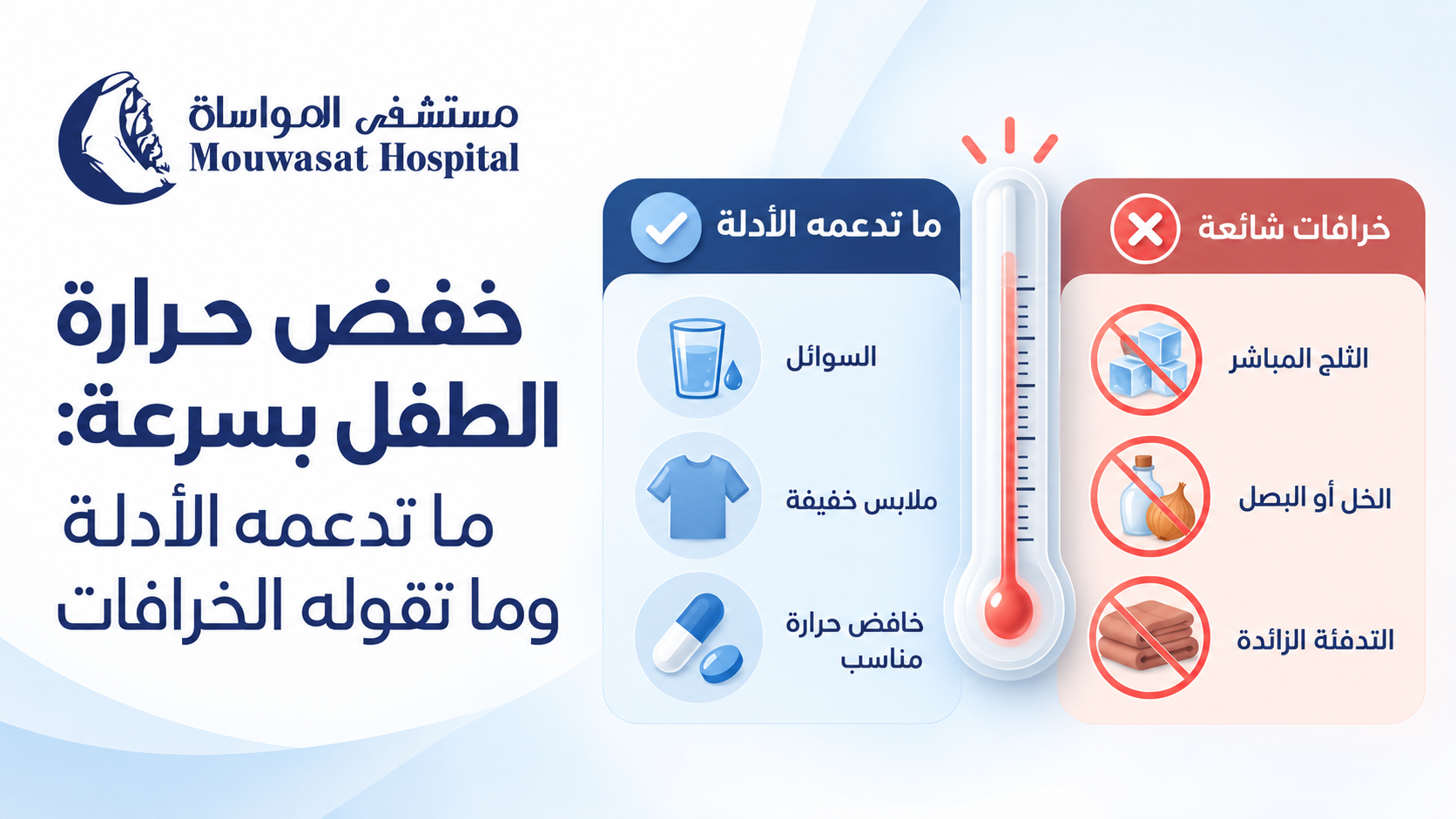
When a child's temperature rises, parents understandably want to know how to bring the fever down quickly. The safest approach, however, is not to force the thermometer back to 37°C as fast as possible.
Good fever care starts by asking whether the child appears well enough for home care or has warning signs that require medical assessment. The main goals are to keep the child comfortable, maintain hydration and monitor their overall condition. A fever falling after medicine does not prove that the illness is minor, and a fever that does not fall quickly does not by itself mean that the illness is serious. [1]
If your child is stable and has no warning signs, the fastest appropriate response is not an ice bath, cold compresses or a home remedy. Focus first on comfort, hydration and observation.
The NHS recommends regular fluids, continued breastfeeding, monitoring for dehydration and avoiding excessive clothing or bedding during a fever. [2]
Children can lose more fluid when they have a fever. Offer age-appropriate fluids regularly and continue normal breastfeeding or milk feeds in infants.
Watch for warning signs of dehydration such as a dry mouth, reduced tears, fewer wet nappies or reduced urination, and unusually low activity. Both NICE and the American Academy of Pediatrics emphasise hydration during fever care. [1] [3]
Do not wrap your child in heavy blankets in an attempt to make them sweat. At the same time, aggressive undressing is not necessary. Use comfortable clothing appropriate for the room temperature.
NICE advises that children with fever should be neither underdressed nor over-wrapped. [1]
A child with a temperature of 38.5°C who remains alert, drinks and interacts can look very different from another child with a lower temperature who is unusually sleepy, dehydrated or struggling to breathe.
Monitor alertness, breathing, drinking, urination, skin colour and associated symptoms as well as the temperature itself.
If you are unsure whether a reading is actually abnormal, see our guide to normal temperature in children and how the measurement site changes the reading.
NICE recommends considering either paracetamol (acetaminophen) or ibuprofen when a child with fever appears distressed or uncomfortable. Fever medicine should not be used solely to reduce the thermometer reading. [1]
This distinction matters. Fever-reducing medicine can make a child feel better, but it does not necessarily treat the cause of the illness or shorten its duration. [3]
NICE advises against giving both medicines at the same time. Changing from one medicine to the other or alternating them may only be considered in specific circumstances when distress continues or returns before the next dose is due. The NHS also advises parents not to alternate them unless a doctor or nurse recommends it. [1] [2]
Always check whether another cold, pain or fever product already contains the same active ingredient before giving more than one medicine.
Parents often become more worried when a fever remains high or decreases only slightly after medicine.
Avoid two common mistakes:
NICE specifically advises healthcare professionals not to use a decrease—or lack of decrease—in temperature after antipyretic medicine to distinguish serious from non-serious illness. [1]
Instead, look at your child. Are they more comfortable? Are they drinking? Are they alert and interacting? Is their breathing normal? Have any new warning signs appeared?
For a broader guide to interpreting fever by age and symptoms, see child fever treatment by age, temperature and symptoms.
Cooling the skin is traditionally used to bring a fever down, but modern guidelines do not recommend physical cooling as the main treatment for routine fever.
NICE does not recommend tepid sponging for treating fever, and the NHS advises against sponging or undressing a child in an attempt to cool them. [1] [2]
Fever is not simply excess heat sitting on the skin. During an infection, the body's temperature regulation changes. Aggressive external cooling may therefore cause discomfort or shivering without treating the underlying cause.
Not necessarily. A comfortable lukewarm bath may feel pleasant to some children, but it should not be used aggressively to chase a thermometer number. Cold water and ice baths should be avoided because they can cause shivering and discomfort. [3]
Saudi Ministry of Health guidance mentions room-temperature water compresses in the specific context of care after a febrile seizure has stopped. That does not mean compresses are the primary treatment for every childhood fever. [5]
A home remedy can be popular in search results without being proven safe or effective. Fever care should distinguish evidence-supported measures from traditional practices.
| Remedy | What the evidence says | Better approach |
|---|---|---|
| Vinegar compresses | Vinegar compresses should not be used to treat fever. Chemical burns have been reported after using vinegar in an attempt to lower fever. [6] | Avoid vinegar and use evidence-based fever care. |
| Rubbing alcohol | Unsafe. Isopropyl alcohol can be inhaled or absorbed through the skin and can cause poisoning. [7] | Never use rubbing alcohol on a child's body to reduce fever. |
| Lemon | A 2026 study reported a temperature reduction after a lemon juice and coconut oil compress, but it used a one-group pre-test/post-test design without a control group. This is not enough to establish effectiveness or overturn current guideline-based fever care. [8] | Do not treat lemon compresses as an established childhood fever therapy. |
| Onion on the feet | This is not part of recommended fever management in NICE, NHS or AAP guidance. | Do not rely on it or allow it to delay appropriate assessment. |
| Olive oil or other oils on the skin | These are not recommended fever-reduction treatments in the clinical guidelines used for this article. | Do not substitute topical oils for recommended fever care. |
| Ice or very cold water | Cold exposure can cause shivering and discomfort and does not treat the cause of the fever. [3] | Avoid aggressive cooling. |
Repeatedly checking the thermometer and trying to reach a specific number can distract from more important signs such as alertness, breathing and hydration.
Antipyretic medicines are mainly intended to relieve distress and discomfort rather than simply normalise body temperature. [1]
Do not change the dose or timing based on one temperature reading. Follow approved instructions and professional advice.
Some cold, flu and pain products may contain paracetamol or another ingredient already given separately. Always check the label.
Alternating medicines can increase the chance of timing or dosing errors and is not a routine approach for every child with fever. [1] [2]
These practices are either unsupported as routine fever treatment or, in the case of vinegar and rubbing alcohol, can cause direct harm. [6] [7]
A falling temperature does not rule out serious illness. Warning signs should be acted on regardless of the response to fever medicine. [1]
No. This is an important misconception because fear of seizures can lead parents to aggressively cool a child or repeatedly give fever medicine.
NICE states that antipyretic medicines do not prevent febrile seizures and should not be used specifically for that purpose. The American Academy of Pediatrics likewise notes that acetaminophen and ibuprofen may lower fever and improve comfort but do not prevent febrile seizures. [1] [4]
If a seizure occurs, do not try to force medicine into the child's mouth and do not put any object between their teeth. Managing the seizure safely becomes the immediate priority.
See our dedicated guide: febrile seizures in children: what to do in the first few minutes.
Seek medical assessment if:
In children older than 6 months, NICE advises that the height of the temperature alone should not be used to identify serious illness. The child's overall condition and accompanying symptoms remain essential. [1]
If you are trying to reduce your child's fever quickly, do not let speed lead to vinegar, rubbing alcohol, ice baths, extra medicine or unsupervised alternating of medicines.
Start by checking for warning signs. Then focus on fluids, breastfeeding, rest and comfortable clothing. If your child is distressed, an appropriate fever medicine may be used according to approved instructions and professional advice.
Remember that the response to fever medicine is not a test of how serious an illness is. The real goal is a more comfortable, adequately hydrated child whose overall condition is being monitored—not a thermometer forced back to 37°C.
For specialist assessment, visit Pediatrics at Mouwasat Hospital.

Seeing a child have a seizure during a fever can be frightening. In those first moments, however, the priority is not to force the seizure to stop or immediately cool the child's body. The most important actions are to protect the child from injury, place them safely on their side, time the seizure and know when emergency medical help is needed.
Most febrile seizures are short and do not cause long-term harm. Some situations do require urgent medical attention, particularly when a seizure lasts more than 5 minutes, it is the child's first seizure, breathing is difficult, or the child does not recover normally afterwards. [1] [2] [3]
Seek emergency medical help if the seizure lasts more than 5 minutes, this is the child's first seizure, breathing is difficult, or the child does not recover normally afterwards. [1] [2]
If your child starts having a seizure, focus on keeping them safe until the seizure stops or emergency help arrives.
Place your child on the floor or another low, safe surface. Move furniture, hard objects and anything sharp out of the way.
Saudi Ministry of Health guidance and the American Academy of Pediatrics recommend placing the child on their side so saliva or vomit can drain from the mouth rather than enter the airway. [1] [3]
Look at the clock as soon as the seizure begins. During a frightening event, a short seizure can feel much longer than it actually is, so accurate timing is extremely useful for both emergency decisions and medical assessment.
If possible, also observe whether the whole body is involved or only one side, whether another seizure occurs, and how the child behaves afterwards.
Do not hold the child's arms or legs down. Restraining the child will not stop the seizure and may cause injury.
A child cannot swallow their tongue. A spoon, cloth, finger or other object in the mouth can injure the teeth or jaw and may obstruct the airway. [2] [3]
Do not leave the child alone. Observe their breathing and skin colour and continue timing the event until the movements stop.
| Do not | Why? |
|---|---|
| Put anything in the mouth | It can injure the mouth or teeth and interfere with the airway. |
| Hold the child down | Restraint does not stop the seizure and can cause injury. |
| Give food, water or oral medicine | The child may not be able to swallow safely during the seizure or before fully recovering. |
| Place the child in a bath | During the seizure, airway safety and preventing injury matter more than trying to lower the temperature. |
| Automatically perform CPR while the child is convulsing | Most children resume normal breathing after the seizure. If breathing does not return after it stops, follow emergency-service instructions immediately. |
Seek emergency medical help if:
Saudi Ministry of Health seizure first-aid guidance identifies a first seizure, a seizure lasting longer than 5 minutes, breathing or waking difficulties, another seizure shortly afterwards, and injury during a seizure as reasons to seek emergency care. [2]
A child may be sleepy or confused for a while after a seizure. This can occur during the normal post-seizure recovery period.
After the movements stop:
Once your child is fully conscious, fever and discomfort can be managed appropriately. However, reducing the temperature does not reliably prevent another febrile seizure. [4]
For safe fever care after your child has recovered, read: how to reduce a child's fever: what the evidence supports and what the myths claim.
The two numbers answer different questions:
| Time | What it means |
|---|---|
| 5 minutes | An emergency-action threshold. An ongoing seizure beyond 5 minutes requires urgent medical help and may require treatment to stop it. |
| 15 minutes | One medical criterion used to distinguish simple from complex febrile seizures. It is not a time to wait before seeking help. |
A simple febrile seizure is typically generalised, lasts less than 15 minutes and does not recur within 24 hours. A complex febrile seizure may last longer, have focal features or recur during the same 24-hour period. [7]
A febrile seizure is a seizure that occurs in some young children around the time of an illness causing fever. They occur most commonly from about 6 months to 5 years of age, and the seizure may occasionally be the first sign that the child is becoming ill. [1] [3]
A child may temporarily lose consciousness, become stiff, have jerking movements of the arms and legs, roll their eyes or have a brief change in facial colour.
A very high fever is not always required. Febrile seizures can occur early in an illness, which is one reason aggressively chasing the thermometer number does not reliably prevent them.
Although the event can look frightening, short, simple febrile seizures generally do not cause brain damage, paralysis, intellectual disability or long-term neurological problems. [3]
Long, recurrent or focal seizures, or a child who does not recover normally, need urgent medical assessment because further treatment or investigation may be needed.
No. Having a febrile seizure does not mean that a child has epilepsy.
Epilepsy involves a tendency to have seizures that are not explained solely by fever or another temporary trigger, whereas febrile seizures occur in association with a febrile illness in young children.
Children who have experienced febrile seizures have a slightly higher future risk of epilepsy than children who have not, but the large majority do not develop epilepsy. [5]
Yes. Some children will have another febrile seizure during a future infection, but many will not.
Recent guidance from Cambridge University Hospitals reports recurrence in approximately 4 in 10 children who have experienced a febrile convulsion. Recurrence is more likely when the first event happens at a younger age or when there is a family history of seizures. [5]
A recurrence does not mean that the child has developed epilepsy, and it does not mean that parents failed to reduce the fever quickly enough.
This is one of the most common misconceptions about febrile seizures.
Paracetamol or ibuprofen may improve comfort during a fever, but they do not reliably prevent febrile seizures.
NICE specifically states that antipyretic medicines do not prevent febrile convulsions and should not be used specifically for this purpose. The American Academy of Pediatrics gives the same advice. [3] [4]
Do not give fever medicine to a child without fever solely because they have previously had a febrile seizure, and do not use extra doses or shorten dosing intervals in an attempt to prevent a seizure.
Useful details include:
If you are unsure whether the temperature itself is abnormal, see: normal temperature in children by measurement site.
For a broader guide to fever severity by age and symptoms, see: child fever treatment by age, temperature and symptoms.
If your child has a febrile seizure, the most important first-aid steps are to turn them onto their side in a safe place, remove objects that could cause injury, time the seizure, avoid restraining them and never put anything in their mouth.
Seek emergency medical help if the seizure lasts more than 5 minutes, it is the child's first seizure, breathing is difficult, seizures recur, or the child does not recover normally afterwards.
Most simple febrile seizures do not cause brain damage and do not mean that a child has epilepsy. Fever-reducing medicines also do not reliably prevent future febrile seizures.
For specialist assessment and follow-up, visit Pediatrics at Mouwasat Hospital.