


Being told that you need a breast biopsy can be frightening, but a biopsy recommendation does not mean that breast cancer has already been diagnosed. It means that a breast lump or imaging abnormality needs tissue analysis to determine what it actually is.
The American Cancer Society states that a biopsy is the only way to know for certain whether an abnormal breast area contains cancer cells. At the same time, most breast biopsy results are not cancer. [1]
A breast biopsy is a procedure that removes a small sample of breast tissue from an abnormal area so that it can be examined in a laboratory by a pathologist.
The sample may be obtained with a thin needle, a larger hollow core needle or, less commonly, through a surgical biopsy. [1]
In most needle biopsies, the purpose is not to remove the entire abnormality. It is to obtain enough tissue to determine what is happening at a cellular level.
A biopsy may be recommended when clinical examination or breast imaging identifies an abnormality that cannot be confidently classified as benign.
Examples include:
Imaging describes how an area looks. Biopsy answers a different question: what is the tissue actually made of?
No.
A biopsy is recommended because imaging or examination cannot provide a sufficiently certain answer. The American Cancer Society notes that most breast biopsy results are not cancer. [1]
The pathology result might show:
Evidence does not support the common belief that standard breast biopsy causes cancer to spread through the body.
The National Cancer Institute states that the chance that tumour biopsy or surgery will cause cancer to spread to other parts of the body is extremely low. Standard medical techniques are designed to minimise this risk. [2]
Avoiding a necessary biopsy because of this fear can be more harmful, because it may delay diagnosis and appropriate treatment if cancer is present.
Fine needle aspiration uses a very thin needle to withdraw fluid or groups of cells. It can be useful in selected situations but generally provides less tissue than a core needle biopsy. [3]
A core needle biopsy uses a hollow needle to remove several small cylinders of tissue.
The American Cancer Society describes core needle biopsy as often the preferred type of biopsy when breast cancer is suspected because it obtains more tissue than fine needle aspiration without requiring open surgery. [4]
A vacuum-assisted device can obtain multiple tissue samples through a single needle insertion and can be useful for selected imaging abnormalities.
A surgical biopsy removes part or all of an abnormal area through an incision.
Most patients who require breast biopsy can undergo a needle procedure rather than surgery. Surgical biopsy is reserved for situations in which needle sampling is not appropriate or has not provided a sufficiently clear answer. [1]

When an abnormal area cannot be reliably targeted by touch, imaging is used to guide the biopsy needle precisely to it.
| Guidance method | When it may be used |
|---|---|
| Ultrasound | When the mass or abnormality is clearly visible on ultrasound, allowing real-time needle guidance. |
| Stereotactic / mammographic guidance | Particularly useful for microcalcifications or abnormalities visible on mammography but not clearly seen on ultrasound. |
| MRI guidance | Used for abnormalities that are visible on MRI but cannot be adequately targeted with mammography or ultrasound. |
RadiologyInfo notes that stereotactic biopsy is particularly useful for small clusters of calcifications and abnormalities not visible on ultrasound, while MRI-guided biopsy is used for findings best seen on MRI. [5] [6]
Most needle breast biopsies are performed using local anaesthetic.
You may feel a brief sting when the anaesthetic is injected. Once the area is numb, pressure or movement may be more noticeable than sharp pain while samples are taken. [3]
Afterward, temporary soreness, bruising, swelling or minor bleeding at the biopsy site can occur.
A very small tissue marker or clip may be placed at the biopsy site.
Its purpose is to mark the exact area that was sampled so that it can be found on future imaging or during treatment if necessary.
The American Cancer Society notes that the marker is usually not visible or noticeable to the patient, can remain in place, is safe during MRI examinations and does not set off standard metal detectors. [4]
Tell your healthcare team about:
Follow the specific instructions given by your healthcare team.
After a needle biopsy, you may be advised to:
Bruising and mild swelling are common after core needle biopsy and can temporarily make the area appear larger. These usually improve with time. [4]
Contact the healthcare team if you develop persistent bleeding, rapidly increasing swelling, worsening pain, increasing redness or warmth, discharge from the biopsy site or fever.

There is no universal turnaround time for every breast biopsy.
The American Cancer Society notes that results typically take at least several days. Additional testing can make the final pathology report take longer. [1]
If breast cancer is identified, further tests on the tissue may include hormone receptor and HER2 testing, which can add information needed for treatment planning.
| Possible result | What it may mean |
|---|---|
| Benign | No cancer is identified; follow-up may be all that is needed if the pathology explains the imaging finding. |
| Atypical or high-risk lesion | Not necessarily cancer but may require additional assessment, surveillance or excision depending on the diagnosis. |
| In situ disease | Abnormal or malignant cells remain within the structure where they began, such as DCIS. |
| Invasive breast cancer | Cancer cells have invaded surrounding tissue and further characterisation is required to plan treatment. |
If invasive breast cancer is found, the biopsy tissue is commonly tested for estrogen receptors ER, progesterone receptors PR and HER2, among other tests when appropriate. These results can directly influence treatment choices. [7]
A benign pathology result needs to make sense in the context of the imaging and clinical examination.
Core needle biopsy provides a clear answer in most cases, but it can occasionally miss a cancer. The American Cancer Society notes that if the result is inconclusive or the doctor remains concerned, another core biopsy or a more extensive surgical biopsy may be needed. [4]
No.
BI-RADS is the standard system radiologists use to describe breast imaging findings and recommend the next step. Some categories require only routine screening, some call for short-term follow-up, while more suspicious categories are more likely to lead to a biopsy recommendation.
A breast biopsy does not mean that cancer has already been diagnosed. It is the procedure used to obtain tissue when examination and imaging cannot determine the nature of an abnormal area with enough certainty.
Most breast biopsies can be performed with a needle and local anaesthetic rather than surgery. Core needle biopsy is commonly used when breast cancer is suspected because it provides enough tissue for diagnosis and additional laboratory tests.
Current medical evidence also does not support the belief that routine breast biopsy “opens” a tumour or causes cancer to spread. The chance of biopsy causing cancer spread is extremely low.
If cancer is found, the tissue provides information beyond the diagnosis itself, including tumour type, grade, ER and PR receptor status and HER2 status. These findings help guide treatment.
If pathology is benign but does not adequately explain a suspicious imaging finding, further evaluation may still be necessary.
When you receive a mammogram report, you may see a number such as BI-RADS 1, BI-RADS 3 or BI-RADS 4. Without context, the number can sound alarming.
BI-RADS stands for Breast Imaging Reporting and Data System. Radiologists use this standardized system to describe breast imaging findings and recommend the next step, such as routine screening, additional imaging, short-term follow-up or biopsy. [1]
Most importantly, a BI-RADS category is not a breast cancer stage and is not itself a cancer diagnosis. It communicates how concerning an imaging finding appears and what should happen next. [2]
The American College of Radiology developed BI-RADS to standardize the language radiologists use when interpreting breast imaging.
The system is used for mammography, breast ultrasound and breast MRI, not mammography alone. [1]
The system helps communicate three practical pieces of information:
| Category | Meaning | Likelihood of malignancy | Typical next step |
|---|---|---|---|
| 0 | Incomplete assessment | Not assigned | Additional imaging and/or comparison with prior studies |
| 1 | Negative | No suspicious imaging finding | Routine screening as appropriate |
| 2 | Benign | Benign imaging finding | Routine screening |
| 3 | Probably benign | ≤2% | Short-interval imaging follow-up, often around six months |
| 4 | Suspicious | >2% to <95% | Tissue diagnosis/biopsy is generally recommended |
| 5 | Highly suggestive of malignancy | ≥95% | Biopsy and appropriate clinical action |
| 6 | Known biopsy-proven malignancy | Cancer has already been confirmed by tissue diagnosis | Treatment planning or response assessment |
BI-RADS 0 does not mean cancer.
It means the radiologist cannot yet assign a final category because additional information is needed. This may include extra mammographic views, magnification, spot compression, ultrasound or comparison with previous mammograms. [2]
Once the additional evaluation is completed, a final BI-RADS category is usually assigned.
BI-RADS 1 means the imaging examination is negative, with no suspicious mass, architectural distortion or suspicious calcification identified. [2]
For someone without symptoms, routine screening is generally appropriate.
However, a negative imaging assessment does not mean that a new persistent clinical symptom should be ignored. A palpable lump or other ongoing breast change should still be discussed with a clinician.
BI-RADS 2 means a finding is present but has a clearly benign imaging appearance.
Examples can include benign calcifications, cysts, lymph nodes or expected changes following a previous breast procedure. [2]
The difference between BI-RADS 1 and 2 is therefore not that category 2 is more dangerous. BI-RADS 1 is negative, while BI-RADS 2 documents a finding known to be benign.
BI-RADS 3 means probably benign.
The likelihood of cancer is no more than 2%. [1]
Rather than immediately performing a biopsy, radiologists commonly recommend short-term follow-up imaging to confirm that the finding remains stable.
The American Cancer Society notes that repeat imaging is often performed within 6 to 12 months and then periodically until the finding has remained stable for a suitable period, commonly at least two years. [2]
If the finding changes during follow-up, the BI-RADS assessment and recommended management may change.
BI-RADS 4 describes a suspicious imaging finding for which tissue diagnosis is generally recommended.
Because category 4 covers a very wide range of cancer likelihoods, it is commonly divided into three subcategories. [1]
| Category | Level of suspicion | Likelihood of malignancy |
|---|---|---|
| 4A | Low suspicion | >2% to ≤10% |
| 4B | Moderate suspicion | >10% to ≤50% |
| 4C | High suspicion | >50% to <95% |
A BI-RADS 4 result therefore does not mean that cancer has been confirmed.
In BI-RADS 4A, for example, most biopsies will not show cancer. However, the level of suspicion is high enough that tissue sampling is appropriate rather than observation alone.
BI-RADS 5 means the imaging appearance is highly suggestive of malignancy, with a likelihood of cancer of at least 95%. [1]
Biopsy is strongly recommended to confirm the diagnosis and provide tissue for further testing.
BI-RADS 6 is fundamentally different from the preceding categories.
It means that the abnormality has already been proven to be malignant by biopsy. [1]
Imaging classified as BI-RADS 6 may be used to assess a known cancer before treatment, during treatment planning or to evaluate response to neoadjuvant therapy.
BI-RADS 6 therefore does not mean “a more advanced cancer than BI-RADS 5.” It simply indicates that tissue diagnosis has already established malignancy.
Not necessarily.
BI-RADS 4 covers a broad range of probabilities, while BI-RADS 5 has a very high likelihood of malignancy. Neither category substitutes for pathology.
A biopsy may show:
If breast cancer is diagnosed, additional tests such as ER, PR and HER2 assessment may be performed on the tissue to help guide treatment.
A benign result can be reassuring, but the pathology result must also be concordant with the imaging finding.
In other words, the benign diagnosis should reasonably explain the abnormality that prompted the biopsy.
If pathology and imaging do not match, additional tissue sampling or further evaluation may be recommended.
No. The same BI-RADS assessment system is also used for breast ultrasound and breast MRI. [1]
No.
BI-RADS reporting also describes breast density using four categories labelled A through D, but these letters do not represent cancer suspicion. [2]
| Density | General meaning |
|---|---|
| A | Breasts are almost entirely fatty. |
| B | Scattered areas of fibroglandular density. |
| C | Heterogeneously dense. |
| D | Extremely dense. |
A report can therefore say:
Breast Density C + BI-RADS 1
without any contradiction. The letter describes breast density; the BI-RADS number describes the imaging assessment.
No.
BI-RADS is an imaging assessment system. Breast cancer staging—such as Stage I, II, III or IV—is determined only after cancer has been diagnosed and additional information about tumour size, lymph nodes, spread and tumour biology has been gathered.
Look at three parts of the report:
| BI-RADS | General next step |
|---|---|
| 0 | Complete the additional imaging or comparison requested. |
| 1–2 | Continue routine screening unless symptoms require separate evaluation. |
| 3 | Attend the recommended short-interval follow-up. |
| 4 | Discuss the recommended biopsy or tissue diagnosis. |
| 5 | Proceed with tissue diagnosis and appropriate clinical evaluation without unnecessary delay. |
| 6 | Continue treatment planning or response assessment for biopsy-proven cancer. |
BI-RADS 3 does not mean that a benign lesion is “turning into” cancer. It means the imaging appearance is probably benign and carries no more than a 2% likelihood of malignancy. Follow-up is used to confirm stability.
No. BI-RADS 4 means the finding is suspicious enough to justify biopsy. Many BI-RADS 4 findings—particularly category 4A—ultimately prove to be benign.
Yes, but it is uncommon. Category 5 carries at least a 95% likelihood of malignancy, which is why tissue confirmation is strongly recommended.
The number itself does not indicate how dangerous the finding is. BI-RADS 0 simply means the assessment is incomplete and more information is needed before a final category can be assigned.
Yes. Additional imaging, comparison with prior studies or follow-up can change the assessment. For example, an initial BI-RADS 0 result may become BI-RADS 1, 2, 3 or 4 after the work-up is completed.
BI-RADS turns breast imaging findings into a standardized assessment and management recommendation. It is not a breast cancer stage and does not itself diagnose cancer.
BI-RADS 0 means the assessment is incomplete; categories 1 and 2 are reassuring; category 3 is probably benign with no more than a 2% likelihood of malignancy and usually requires short-term follow-up.
BI-RADS 4 is suspicious and generally leads to biopsy, with 4A, 4B and 4C representing progressively higher levels of suspicion. BI-RADS 5 carries at least a 95% likelihood of malignancy but still requires tissue confirmation.
BI-RADS 6 is reserved for a cancer that has already been proven by biopsy.
When reading a report, do not focus only on the number. The radiologist's recommendation for the next step is just as important.

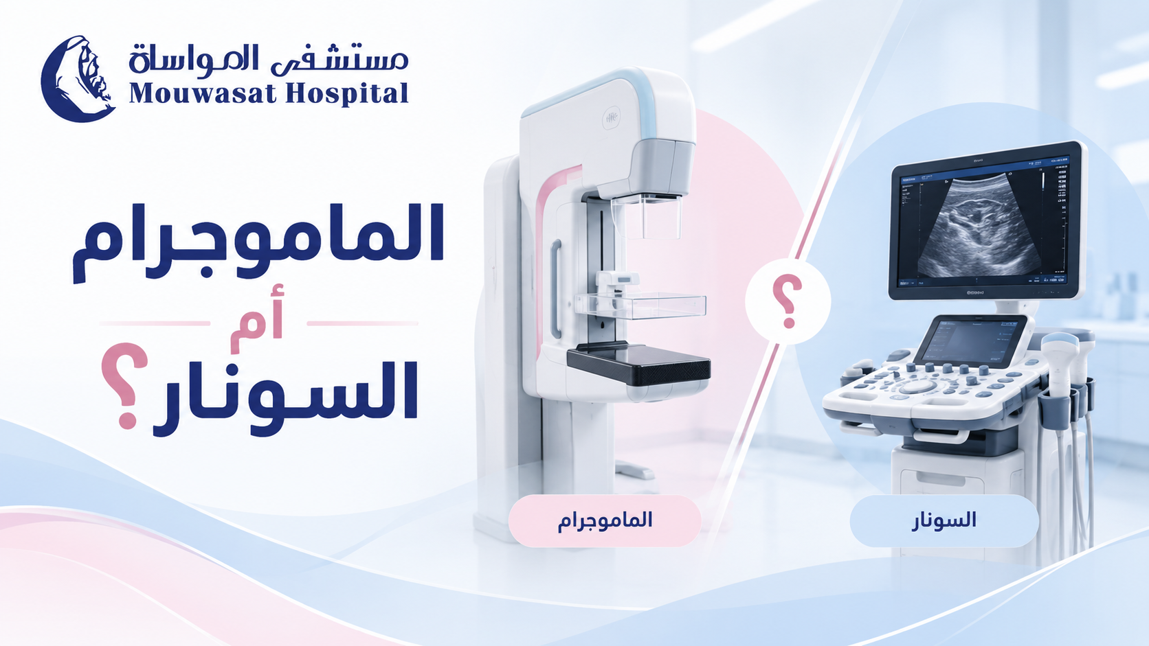
A mammogram and a breast ultrasound are both important breast imaging tests, but they are not interchangeable. Each works differently and answers different clinical questions.
Mammography uses low-dose X-rays and is the primary imaging method used for breast cancer screening. Ultrasound uses sound waves and is particularly useful for evaluating a specific breast lump or abnormality and determining whether a finding is solid or fluid-filled. In many situations, the two tests complement rather than replace each other. [1] [2]
| Feature | Mammogram | Breast ultrasound |
|---|---|---|
| Technology | Low-dose X-rays. | High-frequency sound waves. |
| Main role | Screening and diagnostic breast imaging. | Evaluating focal abnormalities and complementing mammography. |
| Radiation | Uses a low dose of ionising radiation. | Uses no ionising radiation. |
| Microcalcifications | Can detect small calcifications associated with some early breast cancers. | Some calcifications visible on mammography may not be visible on ultrasound. |
| Cysts vs solid masses | May identify a mass or abnormal area. | Very useful for determining whether a finding is fluid-filled or solid. |
The Saudi Ministry of Health describes mammography as low-dose X-ray imaging used for both screening and diagnosis. Breast ultrasound instead produces real-time images using sound waves and does not use radiation. [1] [2]
Mammography is particularly important when the goal is breast cancer screening before symptoms develop.
It can image the entire breast and identify changes that cannot be felt, including small calcifications that may be associated with ductal carcinoma in situ (DCIS) or other breast cancers. [3]
Diagnostic mammography is also used when a patient already has a breast concern, such as:
Breast ultrasound is a safe, non-invasive examination that uses sound waves to evaluate breast tissue. [2]
Common uses include:
| Situation | Typical imaging approach |
|---|---|
| Under 30 with a palpable lump | Breast ultrasound is usually the appropriate first test. |
| Age 30–39 with a new lump | Ultrasound and/or diagnostic mammography or tomosynthesis may be appropriate. |
| Age 40 or older with a new lump | Diagnostic mammography or tomosynthesis is commonly used, often with targeted ultrasound. |
American College of Radiology guidance generally recommends ultrasound first for a palpable mass in women younger than 30, while diagnostic mammography and ultrasound become complementary tools at older ages. [4]
Ultrasound can identify masses and abnormalities that may represent breast cancer, but it cannot detect every breast cancer and cannot always confirm whether a mass is malignant.
A suspicious solid mass may require tissue sampling. A biopsy examines the actual cells and can provide the definitive diagnosis when imaging alone cannot.
No. Radiologists assess characteristics such as shape, margins, orientation and internal appearance, but no single ultrasound image allows a patient to diagnose breast cancer by comparing pictures online.
Yes.
Some early breast cancers, particularly ductal carcinoma in situ (DCIS), may present mainly as small calcifications on mammography without a mass that is visible on ultrasound. This is one reason ultrasound does not replace mammography for breast cancer screening. [2] [3]
Yes. Ultrasound can identify some lesions that are difficult to see clearly on mammography, including some abnormalities in dense breast tissue and findings related to a specific palpable area.
The two tests therefore provide different information rather than competing to determine which is universally “better.”
Breast density refers to the relative amount of fibrous and glandular tissue compared with fatty tissue as seen on a mammogram. It cannot be determined simply by touching the breast. [5]
Dense tissue and many breast abnormalities both appear white on mammograms, which can make some cancers more difficult to detect.
Ultrasound may detect additional findings in women with dense breasts, but supplemental ultrasound also increases false-positive findings and may lead to extra testing or benign biopsies. [2]
No.
More imaging does not always mean better care. Additional ultrasound can identify benign abnormalities that lead to repeat imaging, aspiration or biopsy.
Using both tests is particularly useful when:
One result does not simply cancel out the other.
A palpable lump may not be clearly visible on mammography but can sometimes be characterised with targeted ultrasound. Conversely, suspicious calcifications may be visible on mammography without a corresponding ultrasound abnormality.
If a new palpable lump remains despite reassuring imaging, the clinical finding should still be followed up. Some cancers may have a normal appearance on initial imaging, and biopsy may occasionally be needed when the examination and imaging do not agree. [4]
Imaging can classify a finding as more or less suspicious, but it does not always provide a definitive tissue diagnosis.
Radiologists use the BI-RADS system to describe breast imaging findings and recommend the next step, which may range from routine screening to short-term follow-up, additional imaging or biopsy.
Breast MRI is not a routine replacement for mammography or ultrasound.
For some women at high risk of breast cancer, MRI may be used in addition to mammography because it is more sensitive for detecting certain cancers. [6]
MRI may also be used in selected diagnostic situations, but its greater sensitivity also means that it can detect findings that later prove to be benign.
| Situation | Typical approach |
|---|---|
| No symptoms; routine breast cancer screening | Mammography is the primary imaging test. |
| Palpable lump under age 30 | Ultrasound is usually the first examination. |
| Palpable lump from age 30 onward | Diagnostic mammography and/or ultrasound depending on age and findings. |
| Abnormality on mammography | Targeted ultrasound or additional mammographic views may be recommended. |
| Suspicious microcalcifications | Mammography is particularly important; biopsy may be recommended based on the findings. |
| Dense breasts without symptoms | Mammography remains important; supplemental imaging depends on overall risk. |
| Very high breast cancer risk | MRI may be added to mammography as part of an individualised screening plan. |
The difference between mammography and breast ultrasound is not that one is universally better than the other. They answer different clinical questions.
Mammography is the primary breast cancer screening test and can identify findings such as microcalcifications before a lump can be felt. Ultrasound is especially useful for evaluating a focal area and determining whether a mass is solid or fluid-filled.
Ultrasound therefore does not generally replace mammography, even in women with dense breasts. At the same time, a normal mammogram should not be used to dismiss a new persistent lump; targeted ultrasound or further evaluation may still be appropriate.
The best test is the one that matches the clinical question—and in some situations, the correct answer is to use both.
A mammogram and a breast ultrasound are both important breast imaging tests, but they are not interchangeable. Each works differently and answers different clinical questions.
Mammography uses low-dose X-rays and is the primary imaging method used for breast cancer screening. Ultrasound uses sound waves and is particularly useful for evaluating a specific breast lump or abnormality and determining whether a finding is solid or fluid-filled. In many situations, the two tests complement rather than replace each other. [1] [2]
| Feature | Mammogram | Breast ultrasound |
|---|---|---|
| Technology | Low-dose X-rays. | High-frequency sound waves. |
| Main role | Screening and diagnostic breast imaging. | Evaluating focal abnormalities and complementing mammography. |
| Radiation | Uses a low dose of ionising radiation. | Uses no ionising radiation. |
| Microcalcifications | Can detect small calcifications associated with some early breast cancers. | Some calcifications visible on mammography may not be visible on ultrasound. |
| Cysts vs solid masses | May identify a mass or abnormal area. | Very useful for determining whether a finding is fluid-filled or solid. |
The Saudi Ministry of Health describes mammography as low-dose X-ray imaging used for both screening and diagnosis. Breast ultrasound instead produces real-time images using sound waves and does not use radiation. [1] [2]
Mammography is particularly important when the goal is breast cancer screening before symptoms develop.
It can image the entire breast and identify changes that cannot be felt, including small calcifications that may be associated with ductal carcinoma in situ (DCIS) or other breast cancers. [3]
Diagnostic mammography is also used when a patient already has a breast concern, such as:
Breast ultrasound is a safe, non-invasive examination that uses sound waves to evaluate breast tissue. [2]
Common uses include:
| Situation | Typical imaging approach |
|---|---|
| Under 30 with a palpable lump | Breast ultrasound is usually the appropriate first test. |
| Age 30–39 with a new lump | Ultrasound and/or diagnostic mammography or tomosynthesis may be appropriate. |
| Age 40 or older with a new lump | Diagnostic mammography or tomosynthesis is commonly used, often with targeted ultrasound. |
American College of Radiology guidance generally recommends ultrasound first for a palpable mass in women younger than 30, while diagnostic mammography and ultrasound become complementary tools at older ages. [4]
Ultrasound can identify masses and abnormalities that may represent breast cancer, but it cannot detect every breast cancer and cannot always confirm whether a mass is malignant.
A suspicious solid mass may require tissue sampling. A biopsy examines the actual cells and can provide the definitive diagnosis when imaging alone cannot.
No. Radiologists assess characteristics such as shape, margins, orientation and internal appearance, but no single ultrasound image allows a patient to diagnose breast cancer by comparing pictures online.
Yes.
Some early breast cancers, particularly ductal carcinoma in situ (DCIS), may present mainly as small calcifications on mammography without a mass that is visible on ultrasound. This is one reason ultrasound does not replace mammography for breast cancer screening. [2] [3]
Yes. Ultrasound can identify some lesions that are difficult to see clearly on mammography, including some abnormalities in dense breast tissue and findings related to a specific palpable area.
The two tests therefore provide different information rather than competing to determine which is universally “better.”
Breast density refers to the relative amount of fibrous and glandular tissue compared with fatty tissue as seen on a mammogram. It cannot be determined simply by touching the breast. [5]
Dense tissue and many breast abnormalities both appear white on mammograms, which can make some cancers more difficult to detect.
Ultrasound may detect additional findings in women with dense breasts, but supplemental ultrasound also increases false-positive findings and may lead to extra testing or benign biopsies. [2]
No.
More imaging does not always mean better care. Additional ultrasound can identify benign abnormalities that lead to repeat imaging, aspiration or biopsy.
Using both tests is particularly useful when:
One result does not simply cancel out the other.
A palpable lump may not be clearly visible on mammography but can sometimes be characterised with targeted ultrasound. Conversely, suspicious calcifications may be visible on mammography without a corresponding ultrasound abnormality.
If a new palpable lump remains despite reassuring imaging, the clinical finding should still be followed up. Some cancers may have a normal appearance on initial imaging, and biopsy may occasionally be needed when the examination and imaging do not agree. [4]
Imaging can classify a finding as more or less suspicious, but it does not always provide a definitive tissue diagnosis.
Radiologists use the BI-RADS system to describe breast imaging findings and recommend the next step, which may range from routine screening to short-term follow-up, additional imaging or biopsy.
Breast MRI is not a routine replacement for mammography or ultrasound.
For some women at high risk of breast cancer, MRI may be used in addition to mammography because it is more sensitive for detecting certain cancers. [6]
MRI may also be used in selected diagnostic situations, but its greater sensitivity also means that it can detect findings that later prove to be benign.
| Situation | Typical approach |
|---|---|
| No symptoms; routine breast cancer screening | Mammography is the primary imaging test. |
| Palpable lump under age 30 | Ultrasound is usually the first examination. |
| Palpable lump from age 30 onward | Diagnostic mammography and/or ultrasound depending on age and findings. |
| Abnormality on mammography | Targeted ultrasound or additional mammographic views may be recommended. |
| Suspicious microcalcifications | Mammography is particularly important; biopsy may be recommended based on the findings. |
| Dense breasts without symptoms | Mammography remains important; supplemental imaging depends on overall risk. |
| Very high breast cancer risk | MRI may be added to mammography as part of an individualised screening plan. |
The difference between mammography and breast ultrasound is not that one is universally better than the other. They answer different clinical questions.
Mammography is the primary breast cancer screening test and can identify findings such as microcalcifications before a lump can be felt. Ultrasound is especially useful for evaluating a focal area and determining whether a mass is solid or fluid-filled.
Ultrasound therefore does not generally replace mammography, even in women with dense breasts. At the same time, a normal mammogram should not be used to dismiss a new persistent lump; targeted ultrasound or further evaluation may still be appropriate.
The best test is the one that matches the clinical question—and in some situations, the correct answer is to use both.
When you receive a mammogram report, you may see a number such as BI-RADS 1, BI-RADS 3 or BI-RADS 4. Without context, the number can sound alarming.
BI-RADS stands for Breast Imaging Reporting and Data System. Radiologists use this standardized system to describe breast imaging findings and recommend the next step, such as routine screening, additional imaging, short-term follow-up or biopsy. [1]
Most importantly, a BI-RADS category is not a breast cancer stage and is not itself a cancer diagnosis. It communicates how concerning an imaging finding appears and what should happen next. [2]
The American College of Radiology developed BI-RADS to standardize the language radiologists use when interpreting breast imaging.
The system is used for mammography, breast ultrasound and breast MRI, not mammography alone. [1]
The system helps communicate three practical pieces of information:
| Category | Meaning | Likelihood of malignancy | Typical next step |
|---|---|---|---|
| 0 | Incomplete assessment | Not assigned | Additional imaging and/or comparison with prior studies |
| 1 | Negative | No suspicious imaging finding | Routine screening as appropriate |
| 2 | Benign | Benign imaging finding | Routine screening |
| 3 | Probably benign | ≤2% | Short-interval imaging follow-up, often around six months |
| 4 | Suspicious | >2% to <95% | Tissue diagnosis/biopsy is generally recommended |
| 5 | Highly suggestive of malignancy | ≥95% | Biopsy and appropriate clinical action |
| 6 | Known biopsy-proven malignancy | Cancer has already been confirmed by tissue diagnosis | Treatment planning or response assessment |
BI-RADS 0 does not mean cancer.
It means the radiologist cannot yet assign a final category because additional information is needed. This may include extra mammographic views, magnification, spot compression, ultrasound or comparison with previous mammograms. [2]
Once the additional evaluation is completed, a final BI-RADS category is usually assigned.
BI-RADS 1 means the imaging examination is negative, with no suspicious mass, architectural distortion or suspicious calcification identified. [2]
For someone without symptoms, routine screening is generally appropriate.
However, a negative imaging assessment does not mean that a new persistent clinical symptom should be ignored. A palpable lump or other ongoing breast change should still be discussed with a clinician.
BI-RADS 2 means a finding is present but has a clearly benign imaging appearance.
Examples can include benign calcifications, cysts, lymph nodes or expected changes following a previous breast procedure. [2]
The difference between BI-RADS 1 and 2 is therefore not that category 2 is more dangerous. BI-RADS 1 is negative, while BI-RADS 2 documents a finding known to be benign.
BI-RADS 3 means probably benign.
The likelihood of cancer is no more than 2%. [1]
Rather than immediately performing a biopsy, radiologists commonly recommend short-term follow-up imaging to confirm that the finding remains stable.
The American Cancer Society notes that repeat imaging is often performed within 6 to 12 months and then periodically until the finding has remained stable for a suitable period, commonly at least two years. [2]
If the finding changes during follow-up, the BI-RADS assessment and recommended management may change.
BI-RADS 4 describes a suspicious imaging finding for which tissue diagnosis is generally recommended.
Because category 4 covers a very wide range of cancer likelihoods, it is commonly divided into three subcategories. [1]
| Category | Level of suspicion | Likelihood of malignancy |
|---|---|---|
| 4A | Low suspicion | >2% to ≤10% |
| 4B | Moderate suspicion | >10% to ≤50% |
| 4C | High suspicion | >50% to <95% |
A BI-RADS 4 result therefore does not mean that cancer has been confirmed.
In BI-RADS 4A, for example, most biopsies will not show cancer. However, the level of suspicion is high enough that tissue sampling is appropriate rather than observation alone.
BI-RADS 5 means the imaging appearance is highly suggestive of malignancy, with a likelihood of cancer of at least 95%. [1]
Biopsy is strongly recommended to confirm the diagnosis and provide tissue for further testing.
BI-RADS 6 is fundamentally different from the preceding categories.
It means that the abnormality has already been proven to be malignant by biopsy. [1]
Imaging classified as BI-RADS 6 may be used to assess a known cancer before treatment, during treatment planning or to evaluate response to neoadjuvant therapy.
BI-RADS 6 therefore does not mean “a more advanced cancer than BI-RADS 5.” It simply indicates that tissue diagnosis has already established malignancy.
Not necessarily.
BI-RADS 4 covers a broad range of probabilities, while BI-RADS 5 has a very high likelihood of malignancy. Neither category substitutes for pathology.
A biopsy may show:
If breast cancer is diagnosed, additional tests such as ER, PR and HER2 assessment may be performed on the tissue to help guide treatment.
A benign result can be reassuring, but the pathology result must also be concordant with the imaging finding.
In other words, the benign diagnosis should reasonably explain the abnormality that prompted the biopsy.
If pathology and imaging do not match, additional tissue sampling or further evaluation may be recommended.
No. The same BI-RADS assessment system is also used for breast ultrasound and breast MRI. [1]
No.
BI-RADS reporting also describes breast density using four categories labelled A through D, but these letters do not represent cancer suspicion. [2]
| Density | General meaning |
|---|---|
| A | Breasts are almost entirely fatty. |
| B | Scattered areas of fibroglandular density. |
| C | Heterogeneously dense. |
| D | Extremely dense. |
A report can therefore say:
Breast Density C + BI-RADS 1
without any contradiction. The letter describes breast density; the BI-RADS number describes the imaging assessment.
No.
BI-RADS is an imaging assessment system. Breast cancer staging—such as Stage I, II, III or IV—is determined only after cancer has been diagnosed and additional information about tumour size, lymph nodes, spread and tumour biology has been gathered.
Look at three parts of the report:
| BI-RADS | General next step |
|---|---|
| 0 | Complete the additional imaging or comparison requested. |
| 1–2 | Continue routine screening unless symptoms require separate evaluation. |
| 3 | Attend the recommended short-interval follow-up. |
| 4 | Discuss the recommended biopsy or tissue diagnosis. |
| 5 | Proceed with tissue diagnosis and appropriate clinical evaluation without unnecessary delay. |
| 6 | Continue treatment planning or response assessment for biopsy-proven cancer. |
BI-RADS 3 does not mean that a benign lesion is “turning into” cancer. It means the imaging appearance is probably benign and carries no more than a 2% likelihood of malignancy. Follow-up is used to confirm stability.
No. BI-RADS 4 means the finding is suspicious enough to justify biopsy. Many BI-RADS 4 findings—particularly category 4A—ultimately prove to be benign.
Yes, but it is uncommon. Category 5 carries at least a 95% likelihood of malignancy, which is why tissue confirmation is strongly recommended.
The number itself does not indicate how dangerous the finding is. BI-RADS 0 simply means the assessment is incomplete and more information is needed before a final category can be assigned.
Yes. Additional imaging, comparison with prior studies or follow-up can change the assessment. For example, an initial BI-RADS 0 result may become BI-RADS 1, 2, 3 or 4 after the work-up is completed.
BI-RADS turns breast imaging findings into a standardized assessment and management recommendation. It is not a breast cancer stage and does not itself diagnose cancer.
BI-RADS 0 means the assessment is incomplete; categories 1 and 2 are reassuring; category 3 is probably benign with no more than a 2% likelihood of malignancy and usually requires short-term follow-up.
BI-RADS 4 is suspicious and generally leads to biopsy, with 4A, 4B and 4C representing progressively higher levels of suspicion. BI-RADS 5 carries at least a 95% likelihood of malignancy but still requires tissue confirmation.
BI-RADS 6 is reserved for a cancer that has already been proven by biopsy.
When reading a report, do not focus only on the number. The radiologist's recommendation for the next step is just as important.

After a breast cancer diagnosis, a pathology report may contain terms such as: Invasive Ductal Carcinoma, Grade 2, ER Positive, PR Positive and HER2 Negative. These terms do not all describe the same thing.
The type of breast cancer describes where it began and how it grows. Grade describes how abnormal the cancer cells look and how quickly they may grow. Stage describes the extent of cancer in the body. ER, PR and HER2 are biomarkers that provide information about tumour biology and can directly influence treatment choices. [1] [2]
| Information | Question it answers | Examples |
|---|---|---|
| Type | Where did the cancer start and how does it grow? | Ductal, lobular, inflammatory |
| Biomarkers | Which biological features does the tumour have? | ER, PR, HER2 |
| Grade | How abnormal do the cells look and how quickly might they grow? | Grade 1, 2, 3 |
| Stage | How extensive is the cancer? | Stage 0, I, II, III, IV |
DCIS develops in cells lining the milk ducts but remains within the ducts and has not invaded surrounding breast tissue. It is considered non-invasive or pre-invasive breast cancer and is generally classified as stage 0. [4]
Invasive ductal carcinoma begins in the milk ducts and then grows beyond the duct wall into surrounding breast tissue. It is the most common invasive breast cancer. [3]
The word invasive does not mean that distant metastasis has already occurred. It means the cancer has invaded beyond the tissue where it began.
Invasive lobular carcinoma begins in the breast lobules and spreads into surrounding tissue. It is less common than invasive ductal carcinoma and may have a different growth and imaging pattern. [3]
Inflammatory breast cancer is a rare, fast-growing form of breast cancer in which cancer cells block lymph vessels in the skin. The breast may become red, swollen or develop an orange-peel appearance. [5]
Paget disease is a rare cancer involving the nipple and usually the areola. It may cause redness, scaling, itching or thickening of the nipple and is commonly associated with underlying DCIS or invasive breast cancer. [6]
| Term | Meaning |
|---|---|
| In situ | Abnormal cells remain within the structure where they began and have not invaded surrounding tissue. |
| Invasive | Cancer cells have moved beyond their site of origin into surrounding breast tissue. |
ER stands for estrogen receptor and PR for progesterone receptor.
Some breast cancer cells contain receptors that respond to these hormones. Testing the tumour for ER and PR helps determine whether hormone signalling may be helping drive its growth. [7]
An ER-positive or PR-positive result does not mean that the cancer is “more positive” or more advanced. It identifies a biological feature that can provide an important treatment target.
HER2 is a protein involved in normal breast cell growth. Some breast cancers have increased HER2 activity, causing the cells to grow and divide more rapidly. [7]
HER2 status is important because HER2-positive cancers may respond to medicines designed specifically to target HER2.
IHC testing may be reported as:
The final interpretation should always be taken from the complete pathology report rather than one isolated score. [8]
Triple-negative breast cancer (TNBC) means the cancer cells are negative for:
TNBC accounts for approximately 15% of breast cancers. [9]
No.
It means standard hormone therapies and conventional HER2-targeted therapies do not work through those specific targets. Other treatment approaches, including chemotherapy, immunotherapy and selected targeted treatments, may be appropriate depending on stage and tumour characteristics. [10]
Grade describes how abnormal the cancer cells look under a microscope and gives information about how quickly they are likely to grow and spread. [11]
| Grade | General meaning |
|---|---|
| Grade 1 | Cells resemble normal breast cells more closely and usually grow more slowly. |
| Grade 2 | Intermediate between grades 1 and 3. |
| Grade 3 | Cells look more abnormal and tend to grow and divide faster. |
Grade 3 does not mean Stage III.
Stage describes the extent of cancer and helps guide treatment.
Modern breast cancer prognostic staging combines TNM information with tumour grade and biomarkers such as ER, PR and HER2. [12]
| Stage | Simplified meaning |
|---|---|
| 0 | Non-invasive disease such as DCIS. |
| I | Early invasive breast cancer, generally limited to the breast or with very limited nodal involvement depending on staging factors. |
| II | A larger tumour and/or involvement of nearby lymph nodes without distant metastasis. |
| III | Locally advanced disease involving more regional lymph nodes, breast skin or chest wall, without distant metastasis. |
| IV | Breast cancer that has spread to distant parts of the body. |
M1 indicates distant metastatic disease, whereas M0 means there is no evidence of distant metastasis based on the available assessment. [12]
| Finding | Meaning |
|---|---|
| Invasive ductal carcinoma | The cancer type. |
| Grade 2 | Intermediate tumour grade. |
| ER+ / PR+ | The tumour has hormone receptors that may provide a treatment target. |
| HER2- | The tumour is not classified as HER2-positive based on the final HER2 assessment. |
What this example does not tell us by itself is the final cancer stage. Tumour size, lymph nodes, distant spread and other staging information are still needed.
| Factor | Why it matters |
|---|---|
| Stage | Helps determine the role and sequence of surgery, radiation and systemic treatments. |
| ER / PR | Can make endocrine therapy an important treatment option. |
| HER2 | Can identify tumours that may benefit from HER2-targeted treatment. |
| Triple-negative | Requires treatment strategies that do not rely on hormone receptors or conventional HER2 targeting. |
| Grade | Adds information about tumour biology and expected behaviour. |
A breast cancer report becomes much easier to understand when you separate type, biomarkers, grade and stage.
Type describes where the cancer began and how it grows. ER, PR and HER2 describe tumour biology and can identify important treatment targets. Grade describes how abnormal the cells appear and how quickly they may grow, while stage describes how extensive the disease is.
Triple-negative breast cancer means that ER, PR and HER2 are negative according to the definition of this subtype; it does not automatically mean advanced disease. Likewise, HER2-positive does not mean metastatic cancer, and Grade 3 does not mean Stage III.
The full picture—including tumour type, biomarkers, grade, TNM information, lymph node status and other test results—is what allows the cancer care team to determine the stage and select an appropriate treatment plan.

Breast cancer is far more common in women, but men also have a small amount of breast tissue and can develop breast cancer. Male breast cancer is rare, but the assumption that it “cannot happen to men” can cause some men to ignore a lump or nipple change longer than they should. [1]
The most common warning sign is a new breast lump or thickening, often under or close to the nipple. Other possible signs include nipple or skin changes, nipple discharge and swelling in the underarm area. [2]
Yes.
Everyone is born with some breast tissue. Men typically develop much less breast tissue than women, but the ducts and cells that remain can still develop cancer. [1]
Male breast cancer remains uncommon, but rare does not mean impossible.
One challenge is that many men do not realise they can develop breast cancer. They may assume a lump is caused by fat, muscle, infection or gynecomastia and delay seeking medical attention. [3]
The most common symptom is a new breast lump or thickened area. It is often firm and painless, although not always, and frequently occurs under or near the nipple or areola. [2]
Other possible signs include:
None of these findings proves that cancer is present, but a new or persistent change should be evaluated. [2]
This is an important distinction because gynecomastia is much more common than male breast cancer.
Gynecomastia is an increase in glandular breast tissue in boys or men, often related to changes in the balance between estrogen and testosterone. It may affect one or both breasts and can cause tenderness or discomfort. [4]
However, not every lump around the nipple can be diagnosed as gynecomastia based on touch alone.
| Finding | What it may suggest | What matters |
|---|---|---|
| Diffuse tissue behind the nipple | Can occur with gynecomastia. | A new or unusual change may still need evaluation. |
| Tenderness or pain | Common with gynecomastia. | Pain does not rule out cancer. |
| A discrete firm lump | Can have benign or malignant causes. | It should be examined rather than self-diagnosed. |
| New nipple inversion | Can be a concerning breast change. | Especially when new and one-sided. |
| Nipple discharge | Has several possible causes. | New discharge, particularly if bloody, should be assessed. |
| Skin dimpling | Not typical of simple gynecomastia. | Needs medical evaluation. |
No.
Male breast cancer lumps are often painless, but not always. Gynecomastia, on the other hand, can cause tenderness or pain. [2] [4]
Pain therefore cannot be used as a home diagnostic test. A persistent new lump should be assessed based on the overall clinical picture rather than whether it hurts.
Male breast cancer can occur at any age, although the risk increases with age and most cases are diagnosed in older men. [1]
A younger age therefore does not make breast cancer impossible. A man in his 30s or 40s should not ignore a persistent new breast lump simply because he considers himself too young for the disease.
Risk factors include:
BRCA1 and BRCA2 help repair DNA damage. Certain inherited changes in these genes can increase the risk of several cancers, including breast cancer in men.
Inherited BRCA2 variants are an important risk factor for male breast cancer. [5]
Genetic counselling and testing may be particularly relevant when:
The American Cancer Society advises men with breast cancer, a strong cancer family history or a known inherited mutation in the family to consider genetic counselling and testing. [3]
Not routinely for most men.
The National Cancer Institute states that screening mammography is not usually recommended for men, including many who have increased breast cancer risk. High-risk surveillance should instead be individualized according to family history, inherited risk and medical assessment. [5]
A man who develops a breast lump is in a different situation. Imaging performed because of a symptom is diagnostic evaluation, not routine screening.
Evaluation generally begins with medical history and examination of the breast, nipple and underarm area.
When further assessment is needed, tests may include:
Mammography can be performed in men when there is a breast symptom that needs evaluation. It can help assess masses, breast tissue and suspicious calcifications.
Ultrasound can help characterize an abnormality and can also guide a needle biopsy when needed.
If imaging identifies a suspicious area, a biopsy provides tissue for laboratory analysis and determines whether cancer cells are present. [6]
Being advised to have a biopsy does not itself confirm cancer; the tissue result provides the diagnosis.
If cancer is found, the tissue can be tested for:
These findings, together with the cancer stage, help guide treatment. Most breast cancers in men are hormone-receptor positive. [3]
Cancer arising from the breast ducts is the most common form of male breast cancer, and invasive ductal carcinoma is a major type. [1]
Lobular carcinoma is less common because male breasts generally contain far fewer lobules.
Other rare types can include Paget disease of the nipple and inflammatory breast cancer. [5]
The overall treatment principles are largely similar to those used for breast cancer in women, with adjustments based on the individual patient. Because male breast cancer is uncommon, much of the evidence and treatment approach comes from broader breast cancer research. [7]
Treatment may include:
Treatment is not chosen simply because the patient is male. Stage, ER and PR status, HER2 status, tumour grade, overall health and other tumour characteristics all influence the plan. [7]
Seek medical evaluation for a new or persistent change such as:
There is no need to wait for a lump to become painful or large. Male breast cancer lumps are often painless when discovered. [2]
No. Benign causes, particularly gynecomastia, are much more common. A new, firm or unusual lump should still be evaluated to determine its cause.
Gynecomastia itself is not breast cancer. However, a new lump should not automatically be assumed to be gynecomastia, particularly when it is firm, asymmetric or associated with nipple or skin changes.
Breast pain in men has many possible causes and is often benign. Pain neither confirms nor rules out cancer. A persistent painful area accompanied by a lump or another new change should be assessed.
No. Some cases are associated with inherited genetic changes, particularly BRCA2, but not every male breast cancer is hereditary.
Yes. Family history increases risk but is not required for the disease to occur.
There is no general recommendation for all men to follow a formal monthly self-examination programme. The practical goal is to be familiar with your usual chest and breast area and seek assessment for a new change. Men at increased inherited risk need individualized medical advice.
Yes. Mammography can be used diagnostically in men when needed. This is different from routine mammography screening for men without symptoms.
Male breast cancer is rare, but it does occur. The most common warning sign is a new breast lump, often under or near the nipple.
At the same time, a breast enlargement or lump in a man does not automatically mean cancer; gynecomastia and other benign causes are much more common.
Pain or touch alone cannot reliably distinguish them. A new firm lump, nipple inversion or discharge, skin changes or underarm swelling should be medically evaluated.
Routine mammography is not recommended for most men, but symptoms require diagnostic assessment that may include mammography, ultrasound and biopsy when appropriate.
Family history and inherited BRCA variants—particularly BRCA2—are also important risk factors and may make genetic counselling relevant for the patient and family.

A new breast lump is one of the best-known early signs of breast cancer, but it is not the only possible sign. Some breast cancers can cause skin, nipple or breast-shape changes, while others may cause no noticeable symptoms at an early stage.
It is equally important to remember that a breast lump or change does not automatically mean cancer. Many breast changes are caused by benign conditions, hormonal changes, cysts or infections. A new or unusual change should, however, be medically assessed so its cause can be identified. [1] [3]
Symptoms vary from person to person, and some people have no symptoms at all in the early stages.
According to the Saudi Ministry of Health and CDC, breast changes that deserve attention include:
These symptoms can also occur in conditions that are not cancer. A new or unusual change should nevertheless be checked by a healthcare professional. [1] [2]
No. A new breast lump is the most common symptom, but breast cancer may first appear as a skin or nipple change, breast swelling, a change in shape, or an abnormal finding on imaging.
Mammography can also detect some breast cancers before they become large enough to be felt, so the absence of a lump does not remove the need for appropriate screening. [3]
To learn when screening is recommended, see: breast cancer screening and mammography: when should it start?.
No. A lump cannot be reliably diagnosed by touch alone.
A common belief is that a cancerous lump must always be hard, painless and fixed in place. Although many breast cancers may feel hard or irregular, the American Cancer Society notes that breast cancer lumps can also be round, soft, tender or painful. [3]
At the same time, most breast lumps are not cancer.
For more detail, see: breast lumps: when are they likely to be benign and when should they be checked?.
Sometimes the first noticeable change is in the skin rather than a lump.
Changes that should be assessed include:
The World Health Organization lists changes in breast size or shape, skin dimpling, redness and nipple or skin changes among possible symptoms of breast cancer. [4]
A new change in the nipple can sometimes be a symptom of breast cancer.
Changes may include:
Nipple discharge can also occur with benign conditions, so it is not a diagnosis of cancer by itself. Medical assessment can determine the cause. [2] [5]
Breast pain by itself is not usually a typical symptom of breast cancer. Most breast pain is caused by non-cancerous conditions such as hormonal changes, inflammation or problems involving nearby muscles and tissues.
However, new and persistent pain should not simply be ignored, particularly if it is accompanied by another breast change. The Saudi Ministry of Health includes new persistent breast or nipple pain among changes that warrant attention, while the NHS notes that breast pain alone is not usually a breast cancer symptom. [1] [5]
Yes. A new lump or thickening in the armpit can be a sign that needs medical assessment.
Lymph nodes in the armpit commonly enlarge because of infection or inflammation, but breast cancer can also spread to nearby lymph nodes. In some cases, enlarged lymph nodes may be noticed before a breast tumour is large enough to feel. [2] [3]
Breast redness and swelling can have many causes. Mastitis, for example, can cause redness, warmth, pain and a firm area in the breast, especially during breastfeeding. [6]
A less common form of breast cancer called inflammatory breast cancer can present differently from the typical breast lump. Possible changes include:
Rapid or unusual redness and swelling that does not improve as expected should therefore be assessed, particularly when there is no clear explanation.
Not every breast change is an emergency, but new, persistent or unusual changes should be medically assessed.
| Change | What to do |
|---|---|
| New breast or armpit lump | Arrange a medical assessment rather than trying to diagnose it by touch. |
| New change in breast size or shape | Have it checked if it is unusual for you or persists. |
| New nipple inversion or skin change | Seek medical assessment to identify the cause. |
| Bloody or unusual nipple discharge | Arrange a medical assessment. |
| New persistent breast pain | Have it assessed if it does not settle or occurs with another breast change. |
| Rapid redness, swelling or orange-peel skin | Do not delay assessment, particularly when there is no clear explanation. |
The CDC recommends medical assessment for breast signs or symptoms while emphasising that many such changes are caused by conditions other than cancer. [2]
No. This is important to remember.
The Saudi Ministry of Health and American Cancer Society note that breast changes can be caused by many non-cancerous conditions, including:
Because symptoms can overlap, the purpose of seeing a healthcare professional is not to assume the worst. It is to identify the cause rather than guess. [1] [3]
Breast cancer does not usually develop overnight, but a woman may notice a sign suddenly—for example, finding a lump while showering or noticing a skin change in the mirror.
Cancer can also be present before it becomes visible or large enough to feel. In many cases, “sudden” refers to when the change was first noticed rather than when the disease began.
| Symptoms | Screening |
|---|---|
| A breast change has already been noticed. | Performed in people without symptoms according to age and risk. |
| The change may require diagnostic assessment. | Designed to detect cancer before symptoms appear. |
| Evaluation should not be delayed until the next routine screening date. | Usually follows a recommended mammography schedule based on individual risk. |
If you have a new lump or symptom, the appropriate next step may be a clinical examination and diagnostic imaging rather than simply waiting for your next routine mammogram.
If you have no symptoms, screening remains important because some breast cancers can be detected before any noticeable change develops. [1] [3]
Knowing how your breasts normally look and feel can help you notice a new change, but checking your breasts does not prevent cancer and should not replace recommended mammography or medical evaluation.
Breast awareness helps identify what is unusual for you; determining the cause requires professional assessment. [5]
Breast cancer can also occur in men, although it is much less common. Possible signs include a breast lump, nipple changes or discharge.
Read more in: breast cancer in men: signs that can easily be missed.
A new lump may be one of the first signs of breast cancer, but the earliest noticeable change may also involve the skin, nipple, breast shape, unusual discharge or the lymph nodes under the arm.
At the same time, most breast changes and lumps are not cancer. Finding a change does not mean you have breast cancer; it means that identifying the cause is better than trying to diagnose it from pain, appearance or touch alone.
Screening also remains important even when you feel well because some breast cancers can be detected before symptoms appear.
For a broader overview, see: breast cancer: from early signs to diagnosis, treatment and screening.
For specialist evaluation and care, visit Oncology at Mouwasat Hospital.
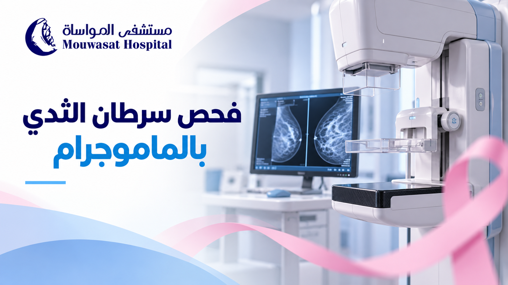
A mammogram is one of the main tools used for breast cancer screening because it can detect breast changes before they become large enough to feel or cause noticeable symptoms. However, mammography is not used for exactly the same purpose in every patient.
There is an important difference between a screening mammogram, which is performed in people without breast symptoms, and a diagnostic mammogram, which is used when there is a lump, nipple discharge, skin change or another finding that needs closer evaluation. [1] [2]
In Saudi Arabia, the Ministry of Health states that mammography is used to screen women aged 40 to 69 years. Women at higher risk may need an earlier or more intensive screening plan based on individual medical assessment. [1]
A mammogram is a low-dose X-ray image of the breast. It can reveal small changes that may not yet be detectable by touch. [2]
Mammography has two main uses:
The Saudi Ministry of Health states that mammography can help identify breast cancer at an early stage before a lesion becomes large enough to be felt. [2]
Saudi Ministry of Health early-detection guidance uses mammography for breast cancer screening in women aged 40 to 69 years. [1]
For a woman around age 40 who has no symptoms and is not known to be at high risk, this is a good time to discuss an appropriate screening schedule with her healthcare provider.
There is no single worldwide interval that applies to every woman. Frequency can depend on age, breast cancer risk, medical history, previous imaging findings and local recommendations.
The safest approach is to follow a screening schedule based on your individual situation rather than selecting an interval from a generic online rule.
| Screening mammogram | Diagnostic mammogram |
|---|---|
| Used when there are no breast symptoms. | Used when there is a symptom or abnormal finding to investigate. |
| Aims to find cancer before symptoms develop. | Aims to study a specific area or concern in greater detail. |
| Usually includes standard views of each breast. | May involve additional views, magnification or other imaging. |
| Part of routine early detection. | Part of a diagnostic work-up and may be combined with ultrasound or other tests. |
Saudi Ministry of Health guidance describes diagnostic mammography as the examination used when a woman has breast symptoms or when a previous mammogram shows a finding requiring further evaluation. [2]
If you have already noticed a change, see: early signs of breast cancer: changes you shouldn't ignore.
Being younger than 40 does not mean mammography is never appropriate. It means that routine population screening is not used in exactly the same way as it is in the main screening age group.
A younger woman with a new breast lump, another breast symptom, a strong family history or other risk factors may need imaging. Depending on the situation, the clinician may use ultrasound, mammography, MRI or a combination of tests.
The more useful question is therefore not simply, “Am I old enough for a mammogram?” but: “Am I symptom-free and considering screening, or do I have a breast problem that needs diagnosis?”
Some women need a personalised screening strategy because their breast cancer risk is significantly higher than average.
Important factors can include:
Saudi guidance notes that breast MRI may be used for women carrying certain genetic mutations associated with increased breast cancer risk. [1]
The American Cancer Society recommends annual mammography plus breast MRI for some women at high risk, typically beginning around age 30, depending on the individual risk profile. [4]
During the examination, you stand in front of the mammography machine. One breast is placed on a special plate and compressed briefly by another plate while an X-ray image is taken.
In a standard screening mammogram, two different views are usually taken of each breast, and the process is then repeated on the other side. [2]
Compression helps:
Saudi Ministry of Health guidance notes that the pressure lasts for a few seconds and does not harm the breast. [2]
Some women feel pressure or discomfort, and some may experience brief pain, particularly when the breasts are already tender.
The compression itself lasts only a short time for each image. The American Cancer Society suggests scheduling the test when the breasts are less swollen or tender if this is an issue for you. [6]
Mammography uses ionising X-rays, so it does involve radiation exposure. However, the radiation dose is low.
The FDA states that the risk of harm from mammography radiation is very small compared with the benefits of early breast cancer detection. [5]
Avoiding an appropriate mammogram because of the belief that the examination itself is more dangerous than an undetected breast cancer therefore does not reflect the balance of current evidence.
There is no evidence that the brief compression used during a mammogram spreads breast cancer. Compression is part of the imaging technique and helps produce a clearer image.
These preparation steps are recommended by the FDA and American Cancer Society to improve image quality and make the examination more comfortable. [5] [6]
Neither test is universally “better” because they are used for different purposes.
Mammography is the main imaging method used for population breast cancer screening, while ultrasound can help assess a specific lump or area and determine whether a mass is solid or fluid-filled. Depending on age, symptoms and breast characteristics, the two tests may be used together. [7]
Read the dedicated guide: mammogram vs breast ultrasound: when is each test used?.
An abnormal mammogram or a request for additional images does not automatically mean breast cancer.
A radiologist may request additional mammographic views, magnification, ultrasound or another examination because an area needs to be assessed more closely.
Breast imaging reports commonly use a standard system called BI-RADS to describe findings and recommend the next step.
See the upcoming guide: mammogram results and BI-RADS: what do categories 0 to 6 mean?.
No screening test is 100% accurate.
Some breast cancers may not be clearly visible on mammography, and mammographic sensitivity can be affected by several factors, particularly breast density. Saudi Ministry of Health information also notes that breast density can influence how well a mammogram detects cancer. [8]
A previous normal mammogram therefore should not be used as a reason to ignore a new breast lump or other new symptom.
No.
Being familiar with the usual appearance and feel of your breasts can help you notice a new change, but it cannot detect every small cancer that may be visible on a mammogram before it becomes palpable.
Not feeling a lump should therefore not be used as a reason to skip recommended breast cancer screening.
| Situation | Typical next step |
|---|---|
| Age 40 or older, no symptoms, average risk | Discuss an appropriate mammography screening schedule based on Saudi guidance and individual assessment. |
| New lump, nipple discharge, skin or nipple change | Diagnostic medical assessment rather than waiting for routine screening. |
| Under 40 with a new breast symptom | Your clinician selects the most appropriate imaging based on age and symptoms. |
| High inherited or family risk | An individualised plan may begin earlier and may include MRI with mammography. |
| Abnormal mammogram result | Additional imaging or other tests as recommended rather than assuming that cancer is present. |
Mammography for breast cancer screening is an important tool for early detection. Saudi Ministry of Health guidance uses mammography to screen women aged 40 to 69, but women at high risk may require a different strategy.
The most important distinction is between screening and diagnosis. If you have no symptoms, mammography may detect breast cancer before you can feel it. If you already have a lump, discharge, nipple change or another new symptom, the appropriate pathway is a diagnostic evaluation rather than simply waiting for routine screening.
Mammography also uses a low radiation dose, and the small radiation risk is outweighed by the benefits of appropriate early detection.
For a broader overview, see: breast cancer: from early signs to diagnosis, treatment and screening.
For specialist evaluation and treatment, visit Oncology at Mouwasat Hospital.