
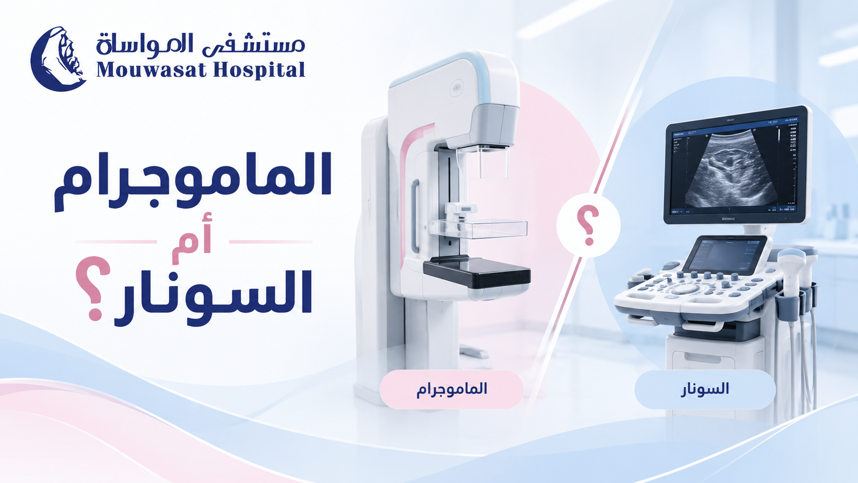

A mammogram and a breast ultrasound are both important breast imaging tests, but they are not interchangeable. Each works differently and answers different clinical questions.
Mammography uses low-dose X-rays and is the primary imaging method used for breast cancer screening. Ultrasound uses sound waves and is particularly useful for evaluating a specific breast lump or abnormality and determining whether a finding is solid or fluid-filled. In many situations, the two tests complement rather than replace each other. [1] [2]
| Feature | Mammogram | Breast ultrasound |
|---|---|---|
| Technology | Low-dose X-rays. | High-frequency sound waves. |
| Main role | Screening and diagnostic breast imaging. | Evaluating focal abnormalities and complementing mammography. |
| Radiation | Uses a low dose of ionising radiation. | Uses no ionising radiation. |
| Microcalcifications | Can detect small calcifications associated with some early breast cancers. | Some calcifications visible on mammography may not be visible on ultrasound. |
| Cysts vs solid masses | May identify a mass or abnormal area. | Very useful for determining whether a finding is fluid-filled or solid. |
The Saudi Ministry of Health describes mammography as low-dose X-ray imaging used for both screening and diagnosis. Breast ultrasound instead produces real-time images using sound waves and does not use radiation. [1] [2]
Mammography is particularly important when the goal is breast cancer screening before symptoms develop.
It can image the entire breast and identify changes that cannot be felt, including small calcifications that may be associated with ductal carcinoma in situ (DCIS) or other breast cancers. [3]
Diagnostic mammography is also used when a patient already has a breast concern, such as:
Breast ultrasound is a safe, non-invasive examination that uses sound waves to evaluate breast tissue. [2]
Common uses include:
| Situation | Typical imaging approach |
|---|---|
| Under 30 with a palpable lump | Breast ultrasound is usually the appropriate first test. |
| Age 30–39 with a new lump | Ultrasound and/or diagnostic mammography or tomosynthesis may be appropriate. |
| Age 40 or older with a new lump | Diagnostic mammography or tomosynthesis is commonly used, often with targeted ultrasound. |
American College of Radiology guidance generally recommends ultrasound first for a palpable mass in women younger than 30, while diagnostic mammography and ultrasound become complementary tools at older ages. [4]

Ultrasound can identify masses and abnormalities that may represent breast cancer, but it cannot detect every breast cancer and cannot always confirm whether a mass is malignant.
A suspicious solid mass may require tissue sampling. A biopsy examines the actual cells and can provide the definitive diagnosis when imaging alone cannot.
No. Radiologists assess characteristics such as shape, margins, orientation and internal appearance, but no single ultrasound image allows a patient to diagnose breast cancer by comparing pictures online.
Yes.
Some early breast cancers, particularly ductal carcinoma in situ (DCIS), may present mainly as small calcifications on mammography without a mass that is visible on ultrasound. This is one reason ultrasound does not replace mammography for breast cancer screening. [2] [3]
Yes. Ultrasound can identify some lesions that are difficult to see clearly on mammography, including some abnormalities in dense breast tissue and findings related to a specific palpable area.
The two tests therefore provide different information rather than competing to determine which is universally “better.”
Breast density refers to the relative amount of fibrous and glandular tissue compared with fatty tissue as seen on a mammogram. It cannot be determined simply by touching the breast. [5]
Dense tissue and many breast abnormalities both appear white on mammograms, which can make some cancers more difficult to detect.
Ultrasound may detect additional findings in women with dense breasts, but supplemental ultrasound also increases false-positive findings and may lead to extra testing or benign biopsies. [2]
No.
More imaging does not always mean better care. Additional ultrasound can identify benign abnormalities that lead to repeat imaging, aspiration or biopsy.
Using both tests is particularly useful when:
One result does not simply cancel out the other.
A palpable lump may not be clearly visible on mammography but can sometimes be characterised with targeted ultrasound. Conversely, suspicious calcifications may be visible on mammography without a corresponding ultrasound abnormality.
If a new palpable lump remains despite reassuring imaging, the clinical finding should still be followed up. Some cancers may have a normal appearance on initial imaging, and biopsy may occasionally be needed when the examination and imaging do not agree. [4]
Imaging can classify a finding as more or less suspicious, but it does not always provide a definitive tissue diagnosis.
Radiologists use the BI-RADS system to describe breast imaging findings and recommend the next step, which may range from routine screening to short-term follow-up, additional imaging or biopsy.
Breast MRI is not a routine replacement for mammography or ultrasound.
For some women at high risk of breast cancer, MRI may be used in addition to mammography because it is more sensitive for detecting certain cancers. [6]
MRI may also be used in selected diagnostic situations, but its greater sensitivity also means that it can detect findings that later prove to be benign.
| Situation | Typical approach |
|---|---|
| No symptoms; routine breast cancer screening | Mammography is the primary imaging test. |
| Palpable lump under age 30 | Ultrasound is usually the first examination. |
| Palpable lump from age 30 onward | Diagnostic mammography and/or ultrasound depending on age and findings. |
| Abnormality on mammography | Targeted ultrasound or additional mammographic views may be recommended. |
| Suspicious microcalcifications | Mammography is particularly important; biopsy may be recommended based on the findings. |
| Dense breasts without symptoms | Mammography remains important; supplemental imaging depends on overall risk. |
| Very high breast cancer risk | MRI may be added to mammography as part of an individualised screening plan. |

The difference between mammography and breast ultrasound is not that one is universally better than the other. They answer different clinical questions.
Mammography is the primary breast cancer screening test and can identify findings such as microcalcifications before a lump can be felt. Ultrasound is especially useful for evaluating a focal area and determining whether a mass is solid or fluid-filled.
Ultrasound therefore does not generally replace mammography, even in women with dense breasts. At the same time, a normal mammogram should not be used to dismiss a new persistent lump; targeted ultrasound or further evaluation may still be appropriate.
The best test is the one that matches the clinical question—and in some situations, the correct answer is to use both.